Echocardiography Simulator
Manual
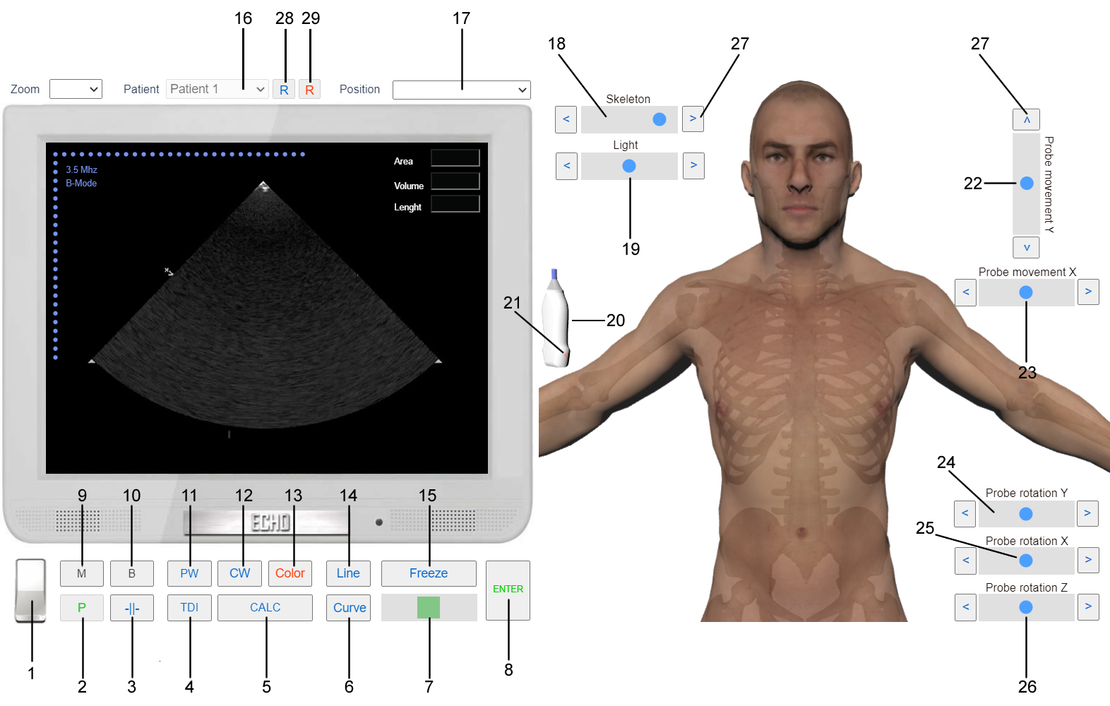
1. Turn on/off the Simulator. 2. Print. 3. Change Zoom, Gain, Contrast, Sephia. 4. Tissue Doppler. 5. Echocardiography Calculations. 6. Curve for Volume and Area measurement. 7. Change slides after freezing the image. 8. Enter Values for Calculation. 9. M-mode. 10. B-mode. 11. Pulse Wave Doppler. 12. Continuous Wave Doppler. 13. Color Doppler. 14. Line for linear measurements. 15. Freeze the image. 16. Change Patient. 17. Change Echocardiography Position. 18. Slider for show/hide sceleton. 19. Slider to increase/decrease light. 20. Transducer. 21. Transducer Marker (Pointer). 22. Slider for Probe movement in Y axis. 23. Slider for Probe movement in X axis. 24. Slider for Probe rotation around Y axis. 25. Slider for Probe tilting around Y axis. 26. Slider for Probe tilting around Z axis. 27. Slider Button. 28. Echocardiography report (User). 29. Echocardiography report (MyEchocardiography.com experts).
Echocadiography Modes and Examinations
- Two-dimensional echocardiography (B-Mode)
- Left Parasternal View. Short axis at the level of Ao Valve
- Left Parasternal View. Short axis at the level of Pulmonary Artery
- Left Parasternal View. Short axis at the level of Mitral Valve
- Left Parasternal View. Short axis at the level of Papillary Muscles
- Left Parasternal View. Short axis at the level of Heart Apex
- Subcostal 4 Chamber View
- Subcostal View of the Inferior Vena Cava
- Subcostal View of the Abdominal Aorta
- One-dimensional echocardiography (M-mode)
- Pulse Wave spectral Doppler (PW). Continuous Wave spectral Doppler (CW)
- Transmitral diastolic flow Spectral Doppler
- Trans-Tricuspid diastolic flow Spectral Doppler
- Pulmonary vein flow Spectral Doppler
- Left ventricular outflow tract (LVOT) flow Spectral Doppler
- Ascending aortic flow Spectral Doppler
- Right ventricular outflow tract (RVOT) flow Spectral Doppler
- Pulmonary artery flow Spectral Doppler
- Inferior vena cava (IVC) flow Spectral Doppler
- Descending aortic flow Spectral Doppler
- Color Doppler
- Transmitral diastolic flow Color Doppler
- Trans-tricuspid flow Color doppler
- Left Ventricular outflow Tract (LVOT) Color Doppler
- Aortic Flow Color Doppler
- Color Doppler of the flow in the Right Ventricular Outflow Tract (RVOT) and Pulmonary Artery
- Color Doppler of the flow in Hepatic Veins and Inferior Vena Cava
Echocardiography Measurements and Calculations
- Linear measurments
- Linear measurements of the left ventricle
- Linear measurements of the right ventricle
- Linear measurements of the Mitral and Tricuspid Annulus
- Linear measurements of the Left and Right Ventricle Outflow Tract
- Linear measurements of the left and right atrium
- Linear measurements of the Aorta
- Linear measurements of the Pulmonary Artery
- Linear Measurement of the Inferior Vena Cava and Hepatic Veins
Systolic function
- LV EF% - Left Ventricle Ejection Fraction (Simpson’s Single Plane, Biplane)
- LV FS% - Left Ventricle Fractional Shortening
- CO - Cardiac Output
- SV - Stroke Volume
- Cl - Cardiac Index
- Local Systolic Function
Diastolic Dysfunction
Assesment severity of the Valvular Pathology
- Ao Regurgitation
- PHT
- Regurgitation jet width / LVOT diameter
- Regurgitation jet area / CSA LVOT
- Vena Contracta
- Regurgitation volume and fraction (SV method)
- TV stenosis
- TV Regurgitation
- PA stenosis
- PA Regurgitation
- PA preassure measurement
Pericarditis
Cardiomyopathies