From the List <<Positions>> Choose Parasternal Short PM (Left Parasternal View, Short axis of LV at the level of Papillary Muscles) or find same position with 3D Transducer
Click the button <<Freeze>> to freeze image. Using Slider find LV End-Diastolic frame.
Click Button <<Calculations>> Click Tab <<LV>> Click Radio Button <<A1>>
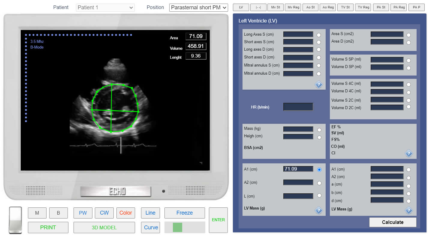
Click the button<<Curve>> place points around epicardium and Click the button <<Enter>>
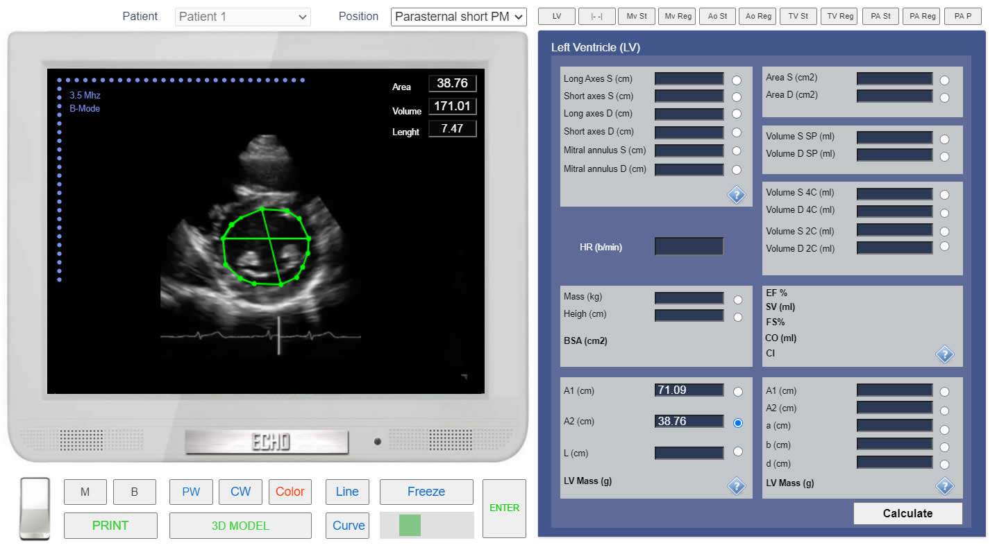
Click Radio Button <<A2>> Click the button<<Curve>> place points around endocardium (ignore papillary muscles) and Click the button <<Enter>>
LV mass. Left parasternal view, at the level of the papillary muscles. Measurement of the Epicardial area (A1).
Click image to enlarge
LV mass. Left parasternal view, at the level of the papillary muscles. Measurement of the Endocardial area (A2).
Click image to enlarge
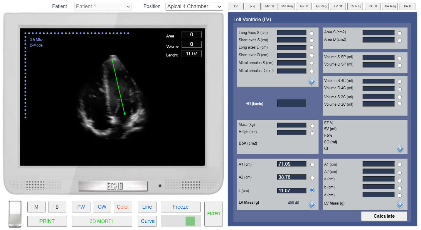
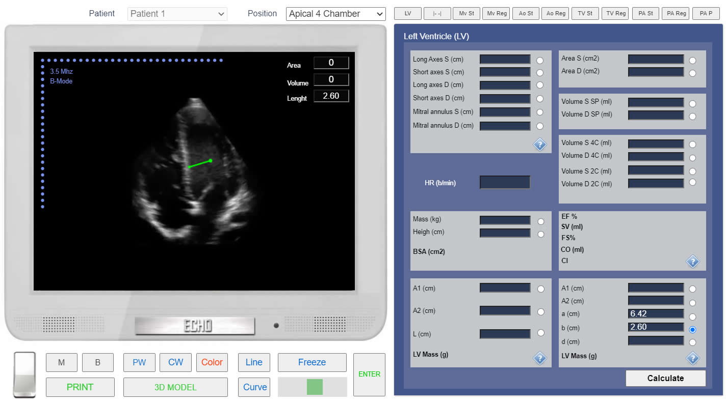
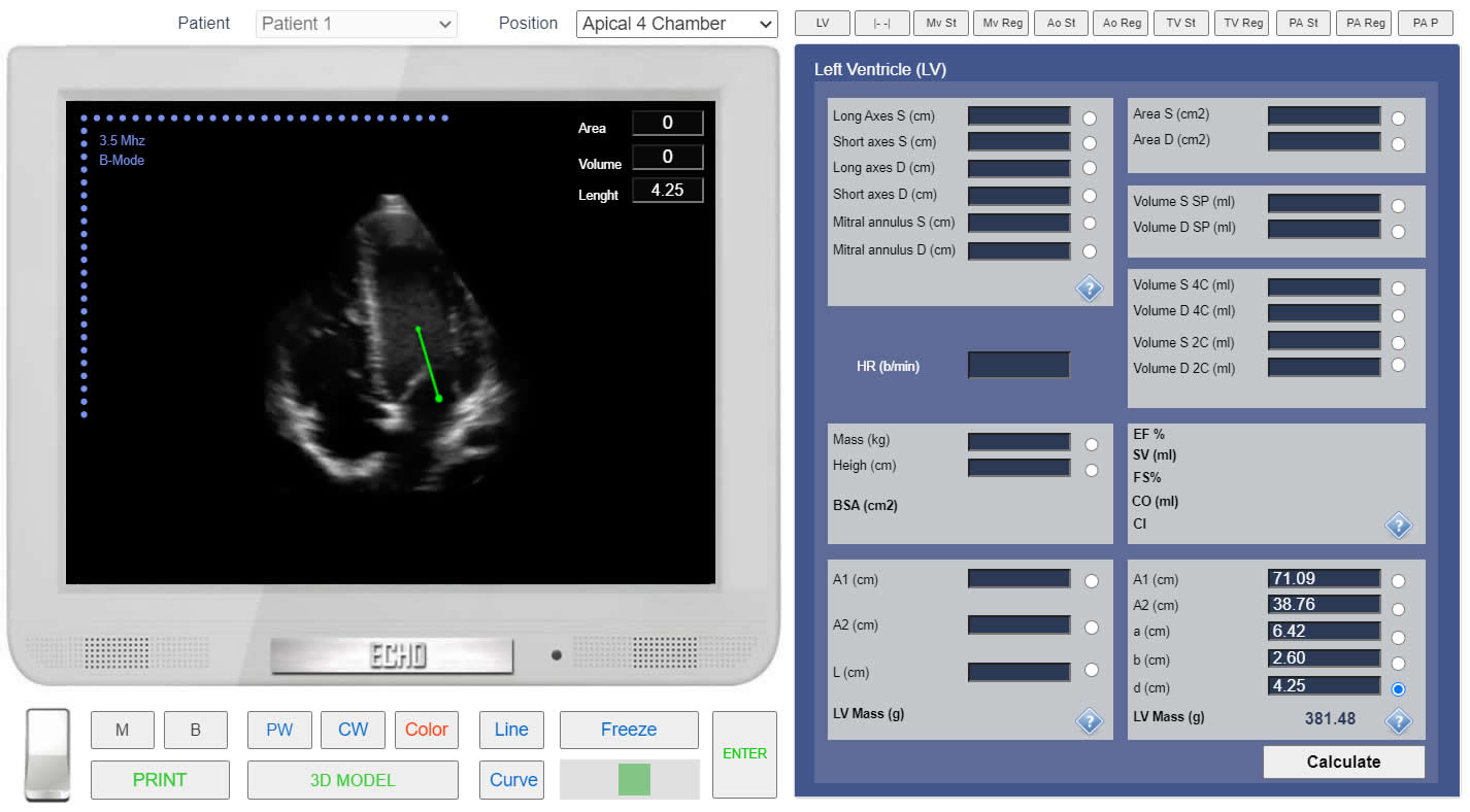
LV mass. Method "Area-Lenght".Apical 4 chamber view. Measurement of the LV Long axis (L).
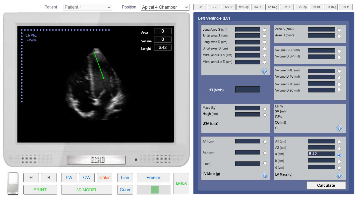
LV mass. Method "Area-Lenght".Simulation By Echocardiography Online Simulator MyEchocardiography.com
Truncated Ellipsoid Method:
PRACTICE IN THE SIMULATOR
From the List <<Positions>> Choose Parasternal Short PM (Left Parasternal View, Short axis of LV at the level of Papillary Muscles) or find same position with 3D Transducer
Click the button <<Freeze>> to freeze image. Using Slider find LV End-Diastolic frame.
Click Button <<Calculations>> Click Tab <<LV>> Click Radio Button <<A1>>
Click the button<<Curve>> place points around epicardium and Click the button <<Enter>>
Click Radio Button <<A2>> Click the button<<Curve>> place points around endocardium (ignore papillary muscles) and Click the button <<Enter>>
 Click image to enlarge
 Click image to enlarge
 Click image to enlarge
 Click image to enlarge
 Click image to enlarge
 Click image to enlarge