left ventricular mass
Echocardiography Textbook
The American Society of Echocardiography (ASE) recommends the use of the following two methods for left ventricular mass assessment (ASE Guideline):
- The "Area-length" method.
- The "Truncated ellipsoid" method.
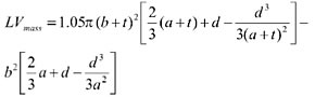
Method "Area-lenght"
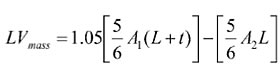
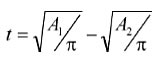
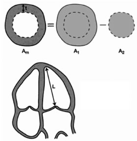
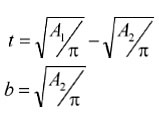
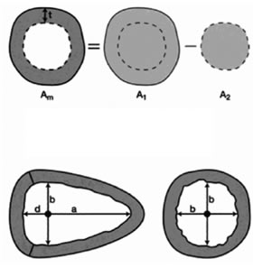
A1 - Epicardial area (cm2). A2 - Endocardial area (cm2). L - Long axis of the left ventricle (cm). t - Mean wall thickness (cm).
In the Left parasternal position, at the level of papillary muscles, contours are drawn around the endocardium and epicardium (at the end of diastole). When tracing the endocardium, do not pay attention to the papillary muscles. Ultrasound devices will automatically calculate the area (A1, A2). The myocardial area is obtained by subtracting the endocardial area from the epicardial.
The long axis of the left ventricle is measured in the Apical four-chamber or two-chamber position, at the end of the diastole.
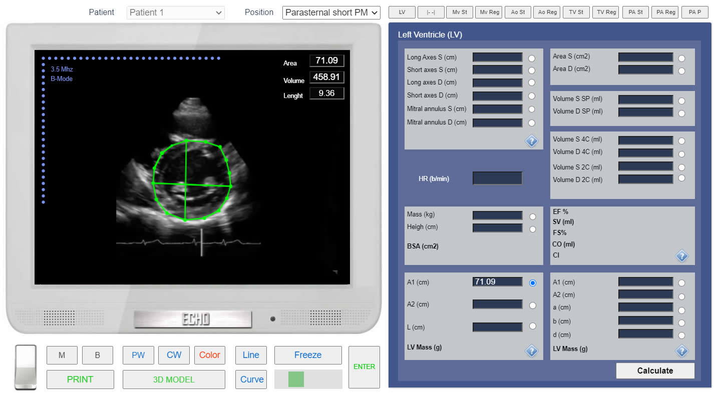
LV mass. Left parasternal view, at the level of the papillary muscles. Measurement of the Epicardial area (A1). Simulation By Echocardiography Online Simulator MyEchocardiography.com
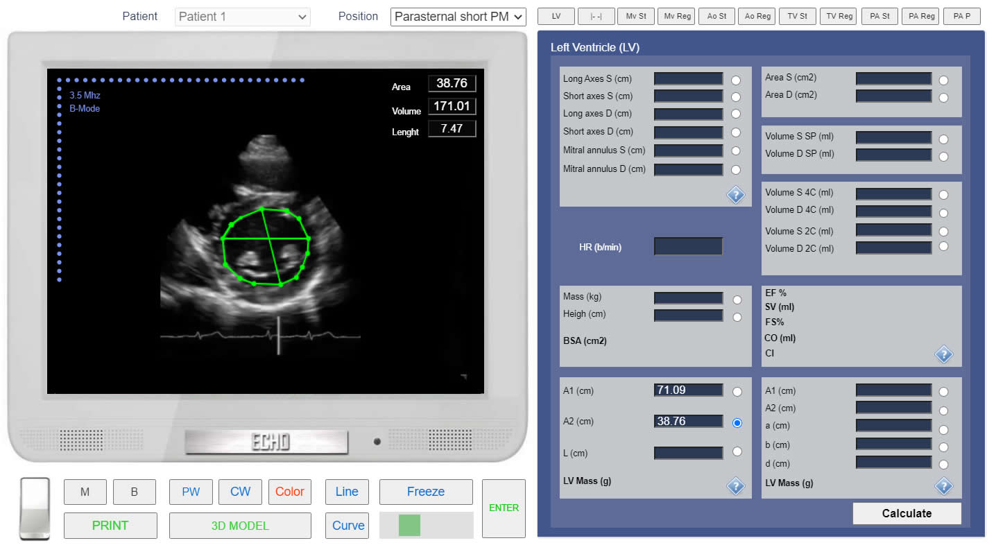
LV mass. Left parasternal view, at the level of the papillary muscles. Measurement of the Endocardial area (A2). Simulation By Echocardiography Online Simulator MyEchocardiography.com
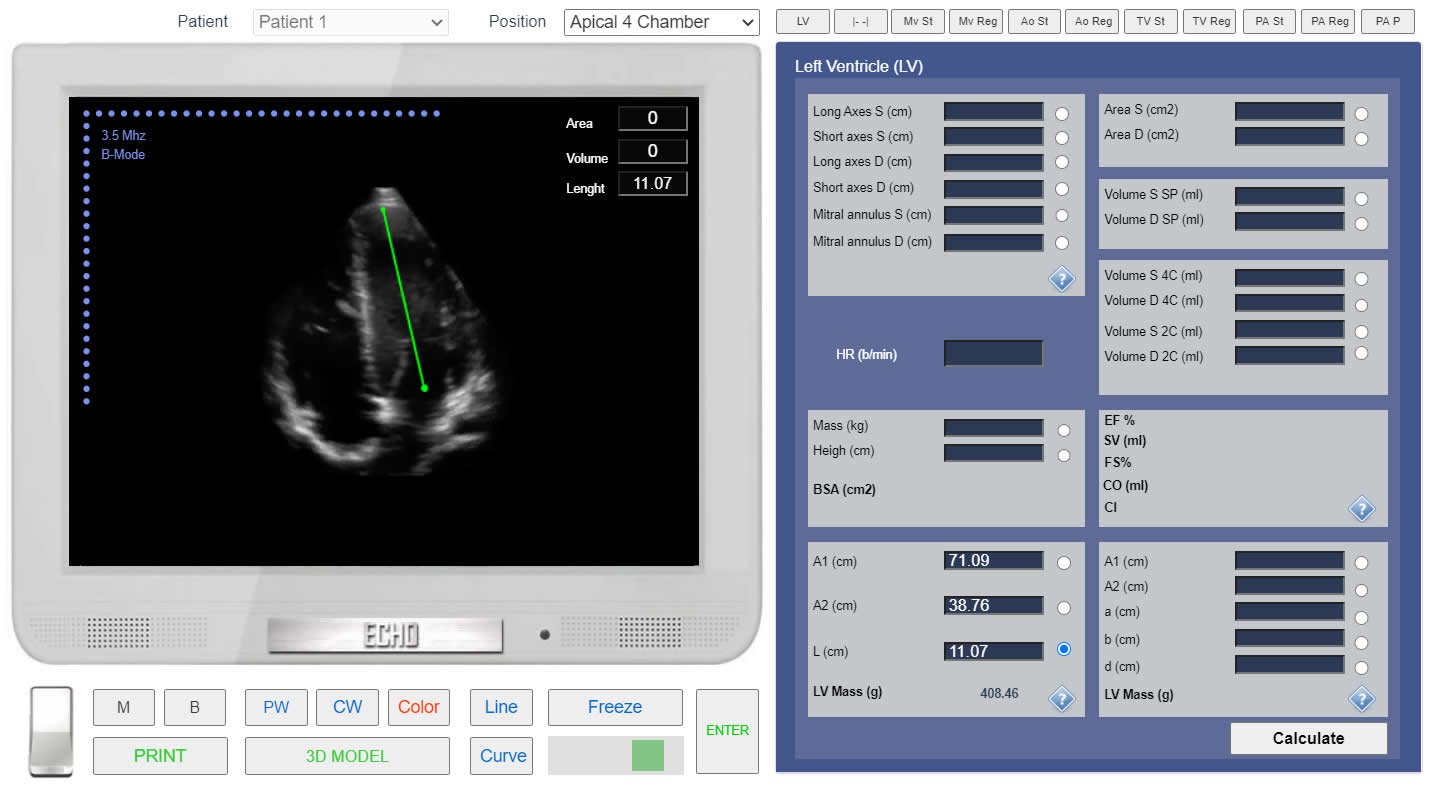
LV mass. Method "Area-Lenght". Apical 4 chamber view. Measurement of the LV Long axis (L). Simulation By Echocardiography Online Simulator MyEchocardiography.com
LV mass. Method "Area-Lenght". Simulation By Echocardiography Online Simulator MyEchocardiography.com
Method "Truncated ellipsoid"
A1 - Epicardial area (cm2). A2 - Endocardial area (cm2). t - Mean wall thickness (cm). a - Long semi-axis of the left ventricle (cm). d - short semi-axis of the left ventricle (cm). b - Short axis radius (cm).
In the Left parasternal position, at the level of papillary muscles, contours are drawn around the endocardium and epicardium (at the end of diastole). When tracing the endocardium, do not pay attention to the papillary muscles. Ultrasound devices will automatically calculate the area (A1, A2). The myocardial area is obtained by subtracting the endocardial area from the epicardial.
Then the left ventricle is presented as a "truncated ellipsoid". The long axis is divided into two parts, at the level of the short axis of the left ventricle. Both parts (a, d) and the radius of the short axis (b) are measured. Measurements have to be done in the Apical four-chamber or two-chamber position, at the end of the diastole.
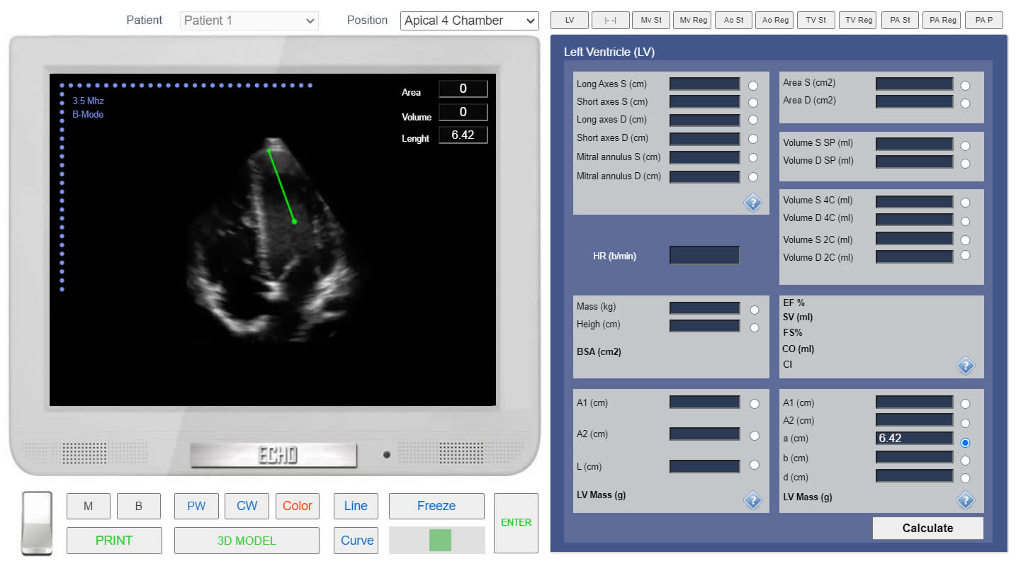
LV mass. Method "Truncated Ellipsoid". Apical 4 chamber view. Measurement of the Long semi-axis of the left ventricle (a). Simulation By Echocardiography Online Simulator MyEchocardiography.com
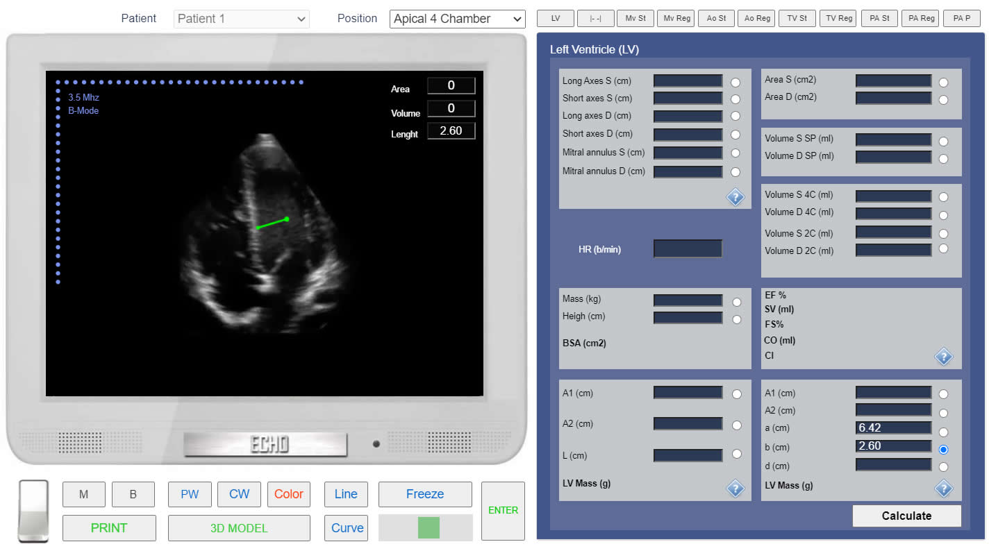
LV mass. Method "Truncated Ellipsoid". Apical 4 chamber view. Measurement of the short axis radius (b). Simulation By Echocardiography Online Simulator MyEchocardiography.com

LV mass. Method "Truncated Ellipsoid". Apical 4 chamber view. Measurement of the short semi-axis of the left ventricle (d). Simulation By Echocardiography Online Simulator MyEchocardiography.com
How to Simulate the LV Mass calculation?