left ventricular Diastolic function
Echocardiography Simulator
What about a little bit of theory before beginning simulation? >>>
From the beginning, the user needs to Calculate LV Ejection Fraction (EF%). Calculations are different in patients with normal EF% than in patients with depressed EF%. See EF% Calculation >>
After Calculating EF% user can start Evaluation of Left Ventricular Diastolic Function:
- From the List <<Patients>> Choose Patient 9 (User can Choose other patients as well).
- Choose 4 chamber position from the List <<Positions>> or find 4 chamber position with 3D Transducer.
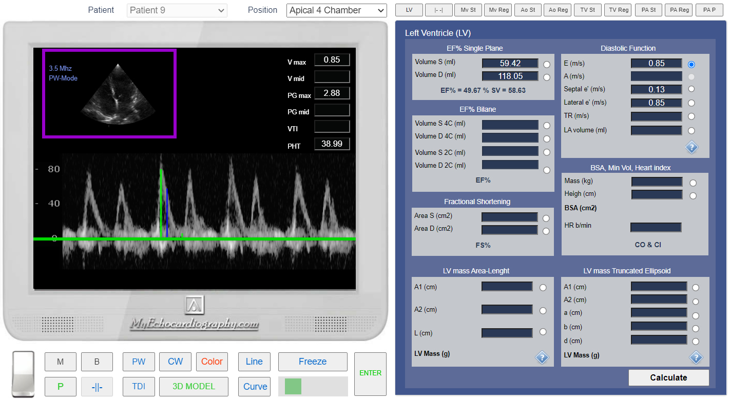
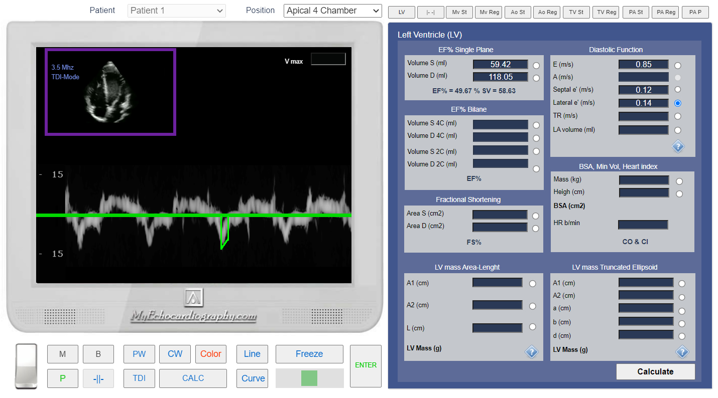
- Click Button <<Calculations>> Click Tab <<LV>> Find Panel "Diastolic Function" and Click Radio Button <<E m/s >>
- Click the button <<PW>> to start Pulse Wave doppler examination. Place Control Volume Beetwen Mitral Valve Leaflets and Click.
- For Mitral Inflow "E" point velocity measurement, click the button <<Line>>. Put first point on the maximum point of the spectrogram. Put another point on baseline.
- Click the button <<Enter>>
* If EF% is depressed, we need to Measure "A" point Velocity as well.
Measurement of the Maximal velocity of trans mitral diastolic flow.
- In the Tab <<LV>> Find Panel "Diastolic Function" and Click Radio Button <<Septal e' m/s >>
- Click The button <<2D>> to move to 2D examination.
- Click the button <<TDI>> to start Tissue doppler examination. Place Control Volume in the septal side of the mitral annulus and Click.
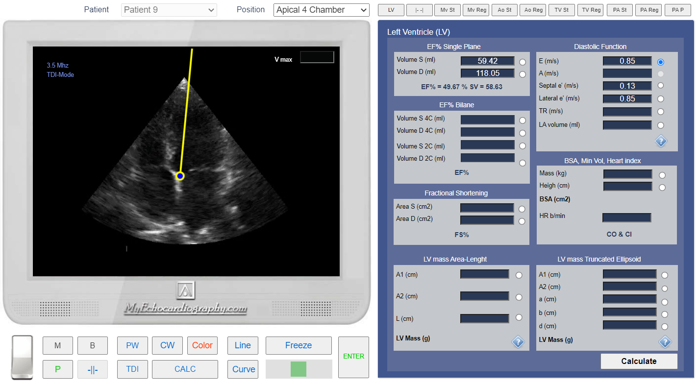
Apical 4 chamber view. Control Volume position for the Septal TDI.
- Click the button <<Line>>. Put first point on the maximum point of the e' vawe. Put another point on Baseline.
- Click the button <<Enter>>
Tissue Doppler (TDI). Measuremen of Septal e' (V max).
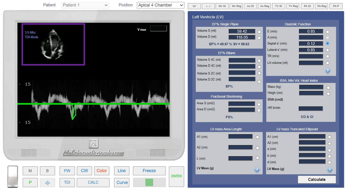
- In the Tab <<LV>> Find Panel "Diastolic Function" and Click Radio Button <<Septal e' m/s >>
- Click The button <<2D>> to move to 2D examination.
- Click the button <<TDI>> to start Tissue doppler examination. Place Control Volume in the lateral side of the mitral annulus and Click.
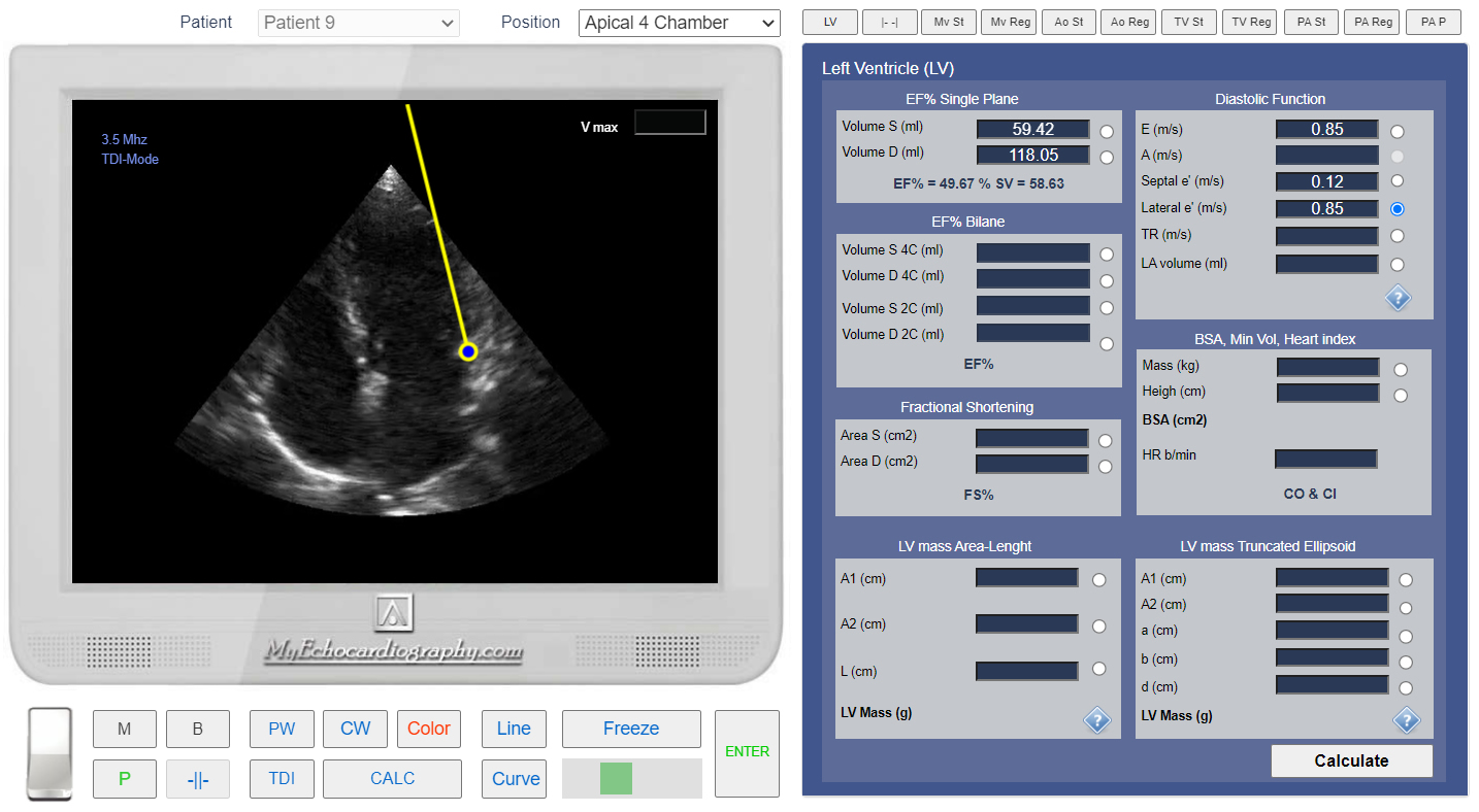
Apical 4 chamber view. Control Volume position for the Lateral TDI.
- Click the button <<Line>>. Put first point on the maximum point of the e' vawe. Put another point on Baseline.
- Click the button <<Enter>>
Tissue Doppler (TDI). Measuremen of the Lateral e' (V max).
- Click the button <<PW>> to start Pulse Wave doppler examination. Place Control Volume beetwen Tricuspid Valve Leaflets and Click.
- Click the button <<Curve>>. Put points around tricuspid regurgitation jet.
- Click the button <<Enter>>
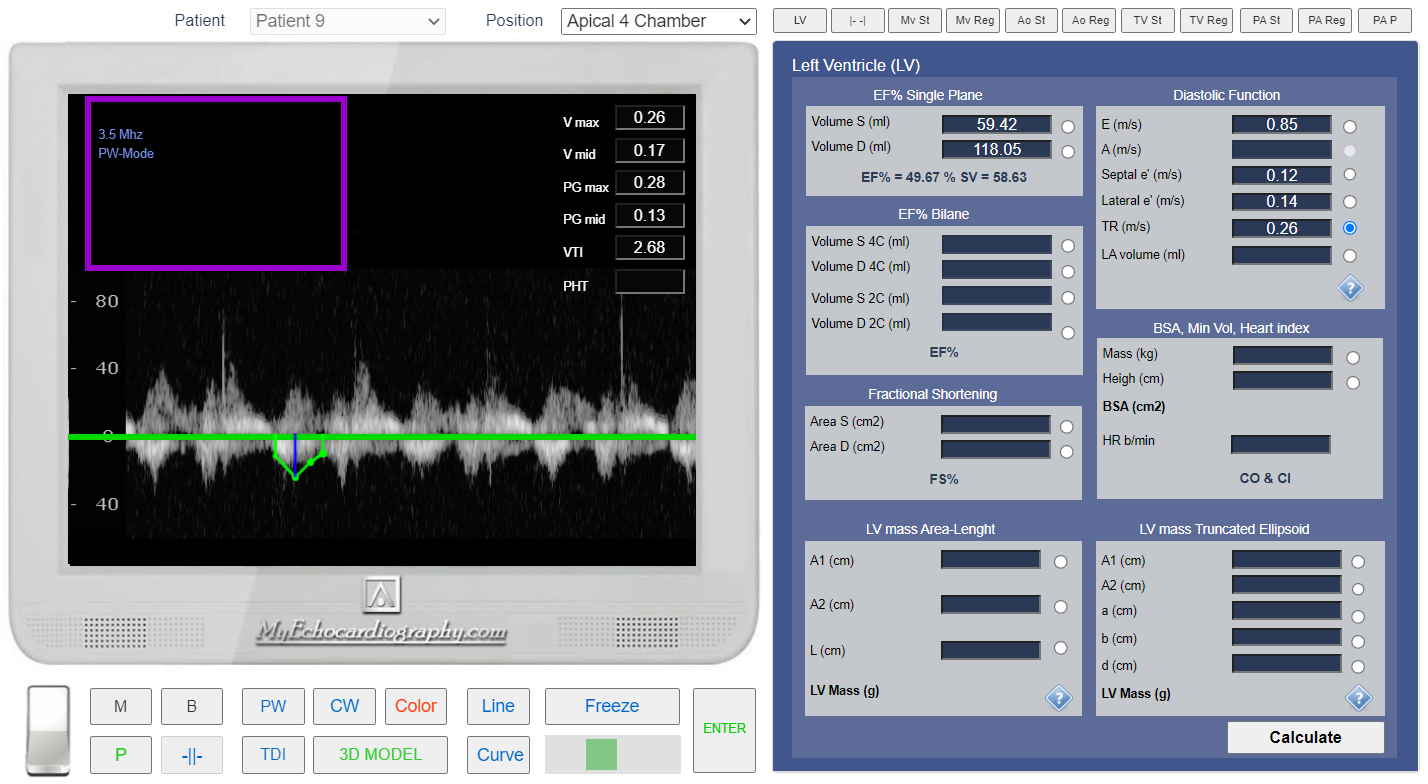
Measurement of the Tricuspid regurgitation velosity.
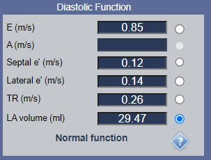
- In the Tab <<LV>>, Find Panel "Diastolic Function" and Click Radio Button <<LA Volume (ml) >>
- Click The button <<2D>> to move to 2D examination.
- Click the button <<Freeze>> to freeze the image. Using Slider find best position for LA Volme measurement.
- Click the button <<Curve>> and trace around inner side of the LA endocardium.
- Click the button <<Enter>>
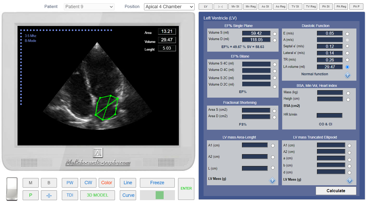
Left Atrium (LA) Volume measurement.
Evaluation of Left Ventricular Diastolic Function with Echocardiography Online Simulator.
Simulation By Echocardiography Online Simulator MyEchocardiography.com