LV Diastolic disfunction
Echocardiography Textbook
According to the guidelines from the American Society of Echocardiography (ASE), There are two algorithms for the evaluation of the left ventricular diastolic function.
First - for the patients with the normal Left Ventricular Ejection Fraction (EF%). Secund - for the patients with depressed EF% and patients with the myocardial disease and normal EF (after consideration of clinical and other 2D data).
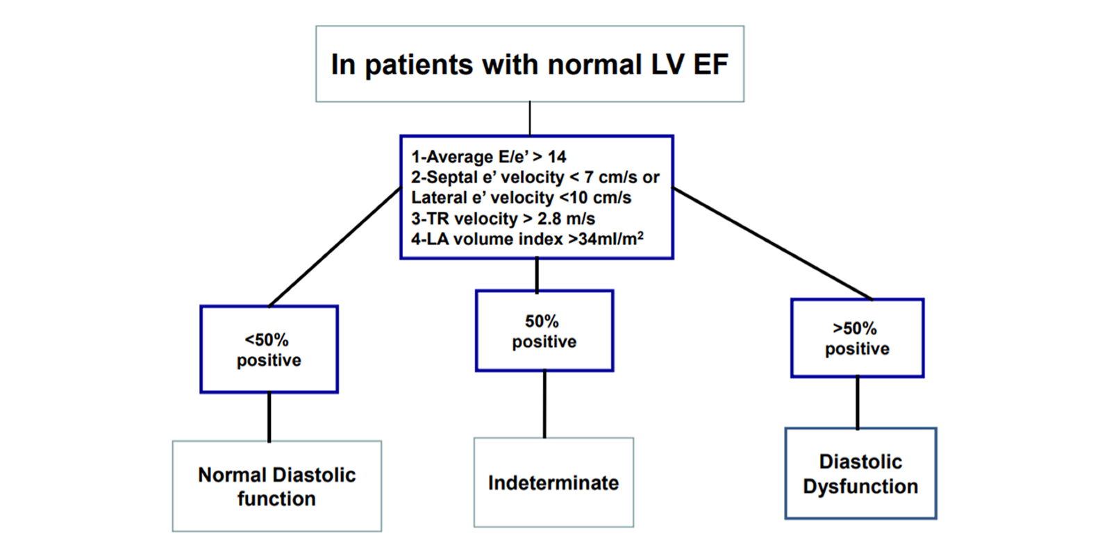
Algorithm for diagnosis of LV diastolic dysfunction in subjects with normal LV EF%.
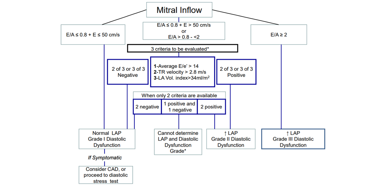
Algorithm for estimation of LV filling pressures and grading LV diastolic function in patients with depressed EF% and patients with myocardial disease and normal EF% (after consideration of clinical and other 2D data).
From the beginning, we need to Calculate LV Ejection Fraction (EF%).
If the LV EF% is normal (and the patient has no myocardial disease) the first algorithm has to be used.
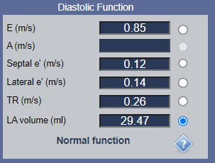
In this case we have to measure 5 parameters.
- E - Maximal velocity of the Transmitral Diastolic Flow (peak E, spectral Doppler).
- Septal e’ - Septal e’ velocity on Tissue Doppler.
- Lateral e’ - Lateral e’ velocity on Tissue Doppler.
- TR velosity - Tricuspid regurgitation velosity.
- LA Volume index - Left Atrial Volume index (for calculation we need LA volume and Body Surface Area - BSA).
If the LV EF% is depressed or EF% is normal, but patients is with myocardial disease the secund algorithm has to be used.
In this case we have to measure 6 parameters.
- E - Maximal velocity of the Early Diastolic Filling (peak E, spectral Doppler).
- A - Maximal velocity of the Late Diastolic Filling (peak A, spectral Doppler).
- Septal e’ - Septal e’ velocity on Tissue Doppler.
- Lateral e’ - Lateral e’ velocity on Tissue Doppler.
- TR velosity - Tricuspid regurgitation velosity.
- LA Volume index - Left Atrial Volume index (for calculation we need LA volume and Body Surface Area - BSA).
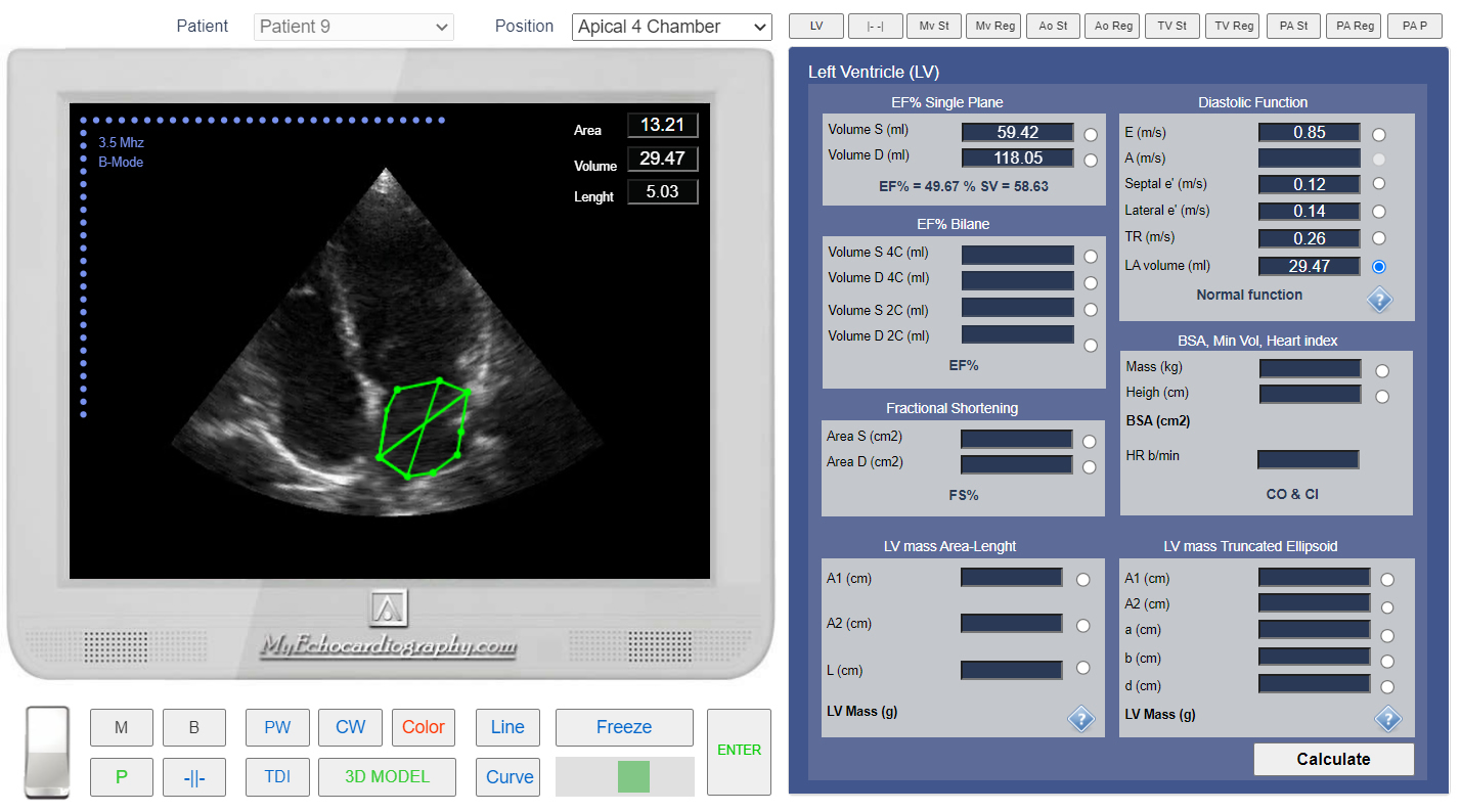
Using Echocardiography Online Simulator MyEchocardiography.com users can simulate all stages of assessment of the LV Diastolic Function.
Simulation of the first algorithm (Normal EF%, no myocardial disease) by Echocardiography Online Simulator:
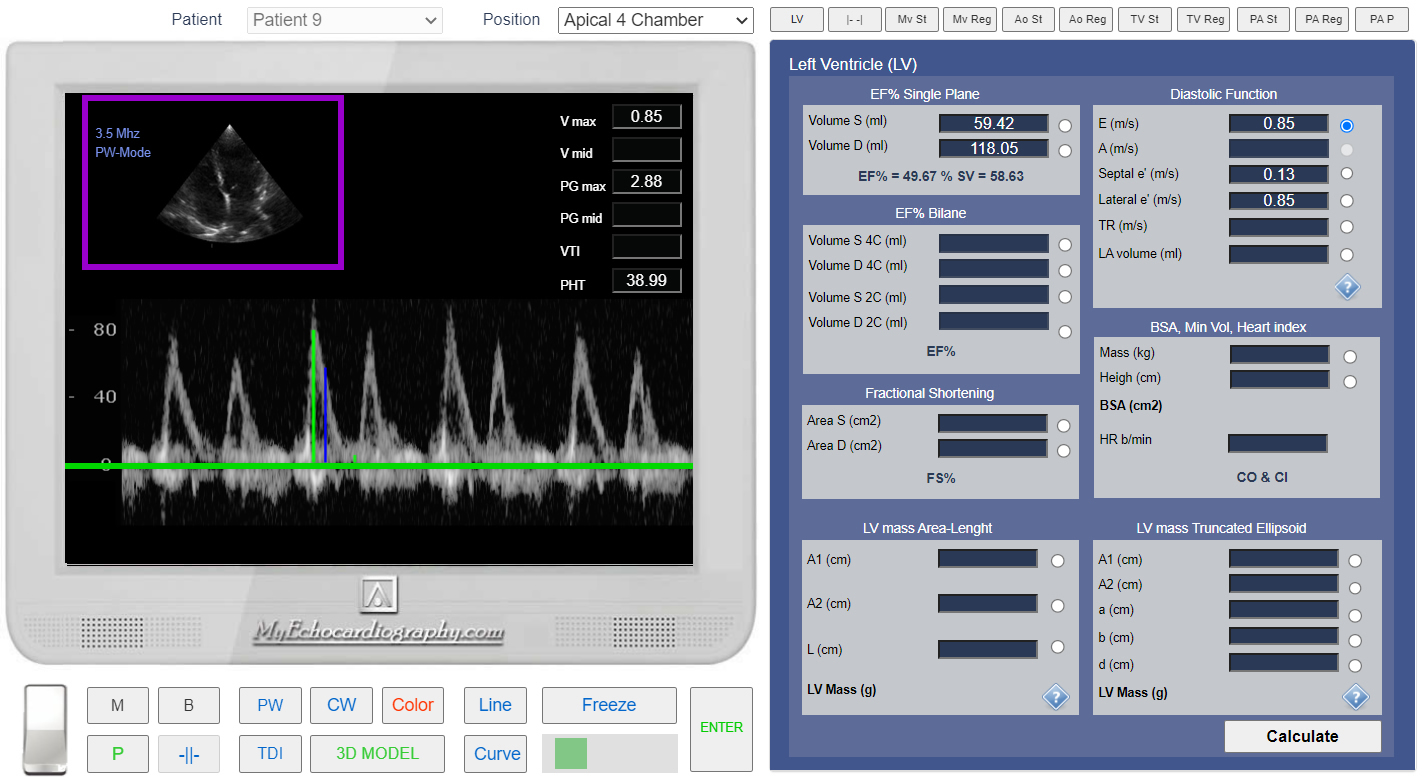
Measurement of the Maximal velocity of trans mitral diastolic flow. Simulation By Echocardiography Online Simulator MyEchocardiography.com
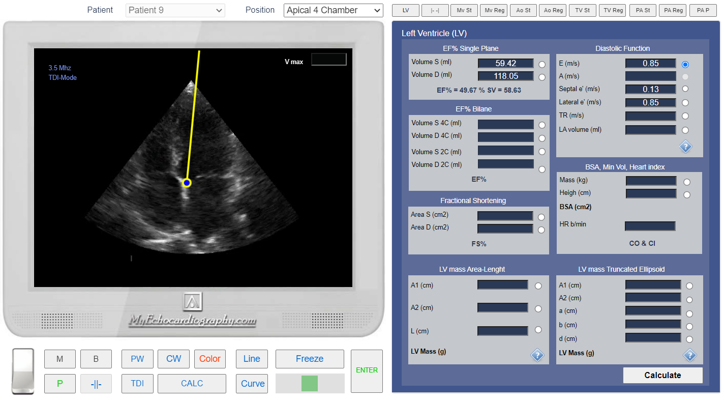
Apical 4 chamber view. Control Volume position for the Septal TDI. Simulation By Echocardiography Online Simulator MyEchocardiography.com
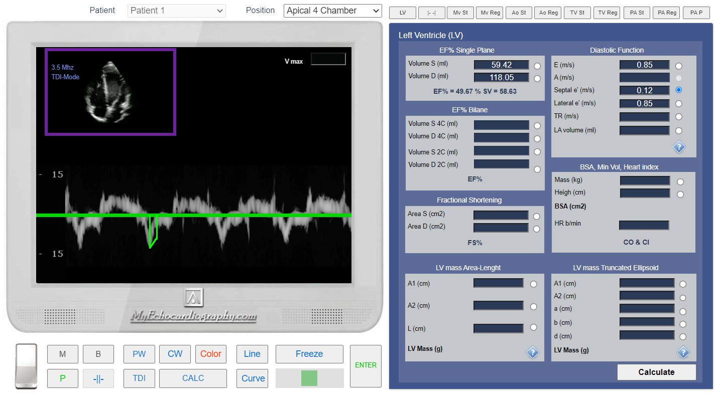
Tissue Doppler (TDI). Measuremen of Septal e' (V max). Simulation By Echocardiography Online Simulator MyEchocardiography.com
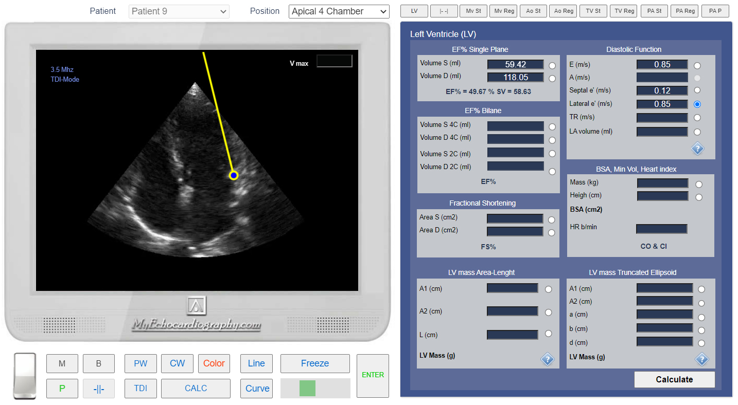
Apical 4 chamber view. Control Volume position for the Lateral TDI. Simulation By Echocardiography Online Simulator MyEchocardiography.com
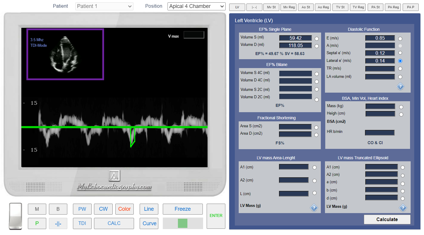
Tissue Doppler (TDI). Measuremen of the Lateral e' (V max).Simulation By Echocardiography Online Simulator MyEchocardiography.com
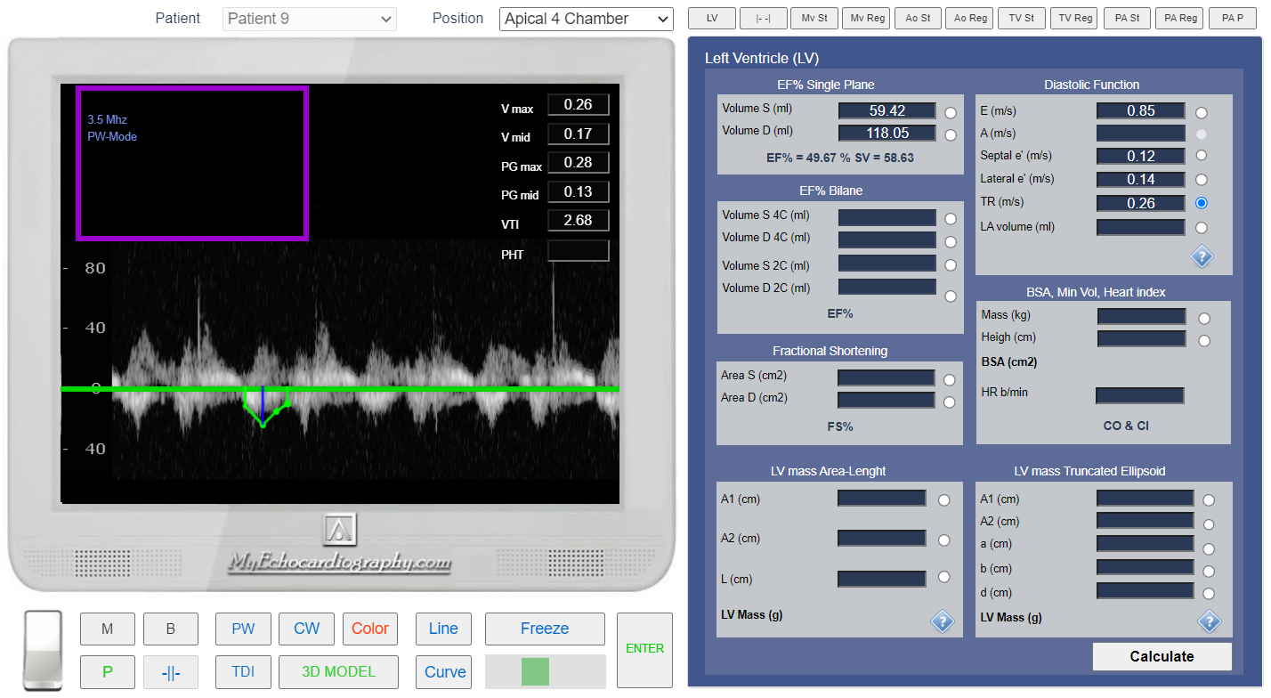
Measurement of the Tricuspid regurgitation velosity. Simulation By Echocardiography Online Simulator MyEchocardiography.com
Left Atrium (LA) Volume measurement. Simulation By Echocardiography Online Simulator MyEchocardiography.com
Evaluation of Left Ventricular Diastolic Function with Echocardiography Online Simulator.
Simulation By Echocardiography Online Simulator MyEchocardiography.com
ASE Recommendations for the Evaluation of Left Ventricular Diastolic Function
How to Simulate Assessment of the Left Ventricular Diastolic Function? >>>