Echocardiographic evidence of mitral regurgitation
Echocardiography Textbook
One and two dimensional Echocardiography
There are no direct echocardiographic signs of mitral regurgitation using 1D and 2D echocardiography.
The only relatively reliable finding can be Incomplete closure of the valve, on a one-dimensional echocardiogram, which is rarely seen.
non-specific symptoms: Increase in the size of the left atrium. LV Myocardial hypertrophy and dilatation.
Color Doppler
The most informative method is color doppler. Regurgitation flow is observed in the systolic phase, which returns to the left atrium (away from the transducer) and is encoded in blue color. Its size and volume depend on the severity of regurgitation.
Slight regurgitation can be identified in 40-60% of healthy people (physiological regurgitation).
The best positions for evaluation: Apical 4 Chamber, Apical 2 Chamber Views, and the Left Parasternal View, long axis.
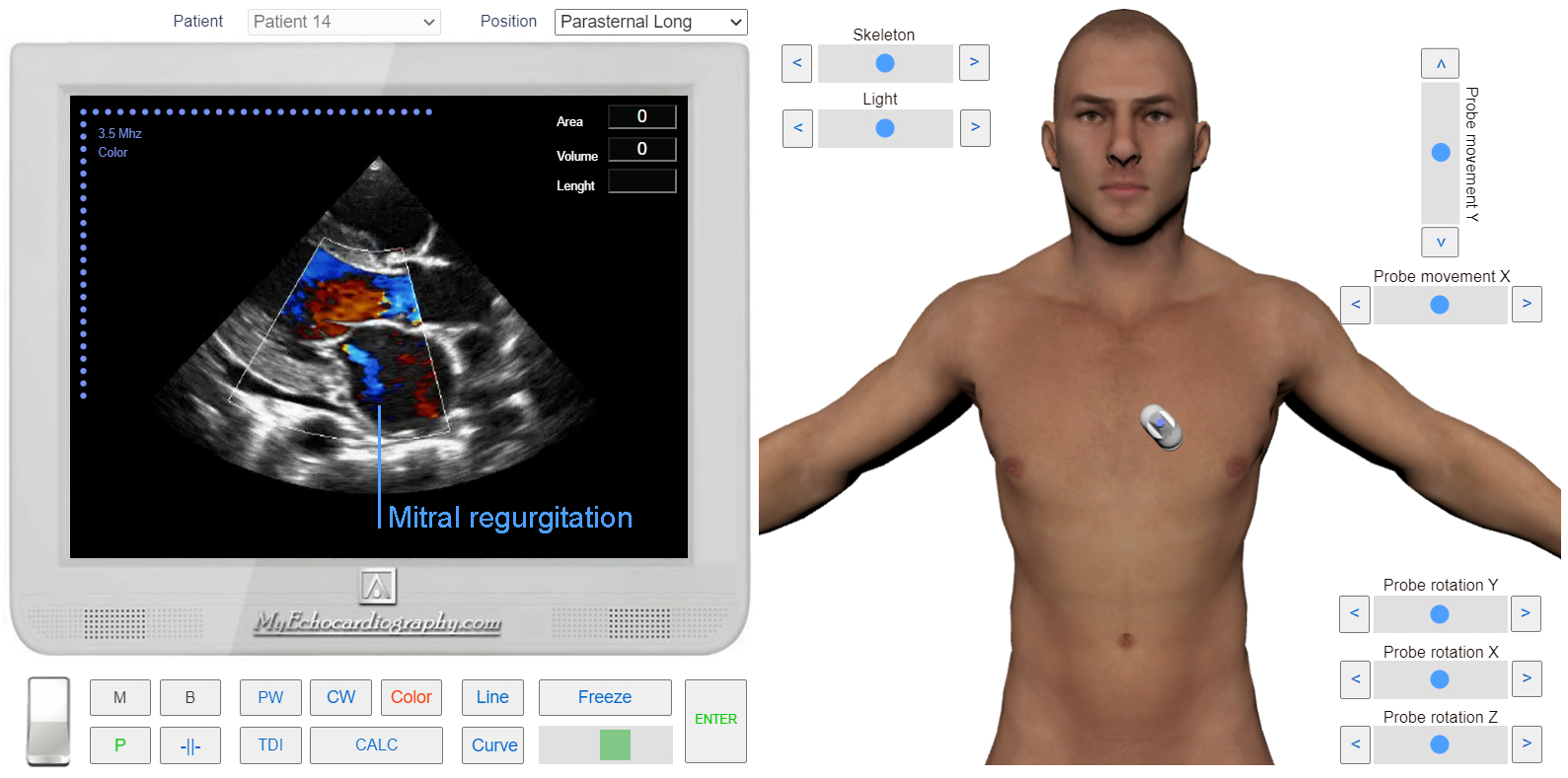
Left Parasternal View, the long axis. Color Doppler. Mitral regurgitation. Simulation by Echocardiography Online Simulator MyEchocardiography.com
Spectral Doppler (PW, CW)
The best positions for evaluation: Apical 4 Chamber, Apical 2 Chamber Views.
To find the regurgitation and determine the depth of its penetration the sample volume has to be moved at different distances from the mitral valve. From the junction of the valves to the upper and lower walls of the left atrium.
The flow of regurgitation is clearly visible on the spectrogram (both in PW and CW mode) as a characteristic spectrum that is directed downward from the baseline. The flow captures the entire systole (from the closing of the mitral valve to the opening of the aortic valve).
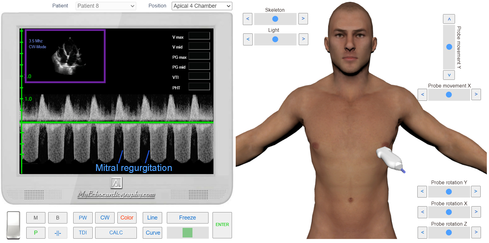
Apical 4 Chamber View. CW Doppler. Mitral regurgitation. Simulation by Echocardiography Online Simulator MyEchocardiography.com
Methods of assessment severity of the mitral Regurgitation:
- Regurgiration Area
- Reg Area / LA area
- PISA
- Regurgitation volume and fraction (CV method)
- Vena Contracta