degree of Mitral Regurgitation with Proximal Isovelocity Surface Area (PISA)
Echocardiography Simulator
What about a little bit of theory before beginning the simulation? >>>
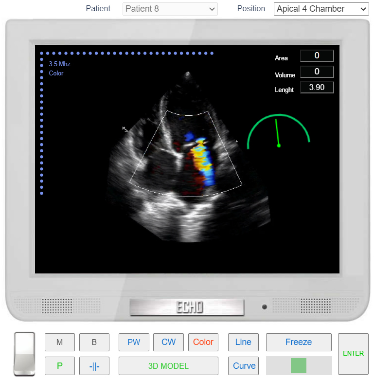
- From the List <<Patients>> Choose Patient 8 (User can Choose other patients with mitral regurgitation as well).
- From the List <<Positions>> Choose Apical 4 Chamber (Apical 4 Chamber view) or find the same position with 3D Transducer.
- Click the button <<Color>>
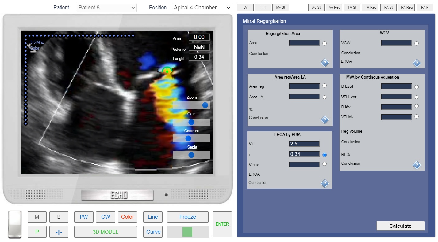
- Click the button <<Freeze>> to freeze image. Click the Button << -||- >> and increase zoom level. Using Slider find the best frame for PISA measerement. The Nyquist Limit is set to 2.5.
- Click Button <<Calculations>> Click Tab <<MV Reg>> Click Radio Button <<r>> (Calculator EROA by PISA).
- Click the button <<Line>> measure radius of the PISA.
- Click the button <<Enter>>.
Apical 4 Chamber view. Color Doppler. Degree of Mitral Regurgitation by Proximal Isovelocity Surface Area (PISA).
Apical 4 Chamber view. Color Doppler. Degree of Mitral Regurgitation by Proximal Isovelocity Surface Area (PISA). Zoomed image.
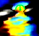
PISA radius (r)
- Click the button <<CW>> to start Continuous Wave doppler examination. Place Control Volume Beetwen Mitral Valve Leaflets and Click.
- In the Tab <<MV Reg>> Click Radio Button <<Vmax>> (Calculator EROA by PISA).
- Click the button <<Curve>> place the points around spectrogram of Mitral Regurgitation. See Spectral Doppler Measurements.
- Click the button <<Enter>>
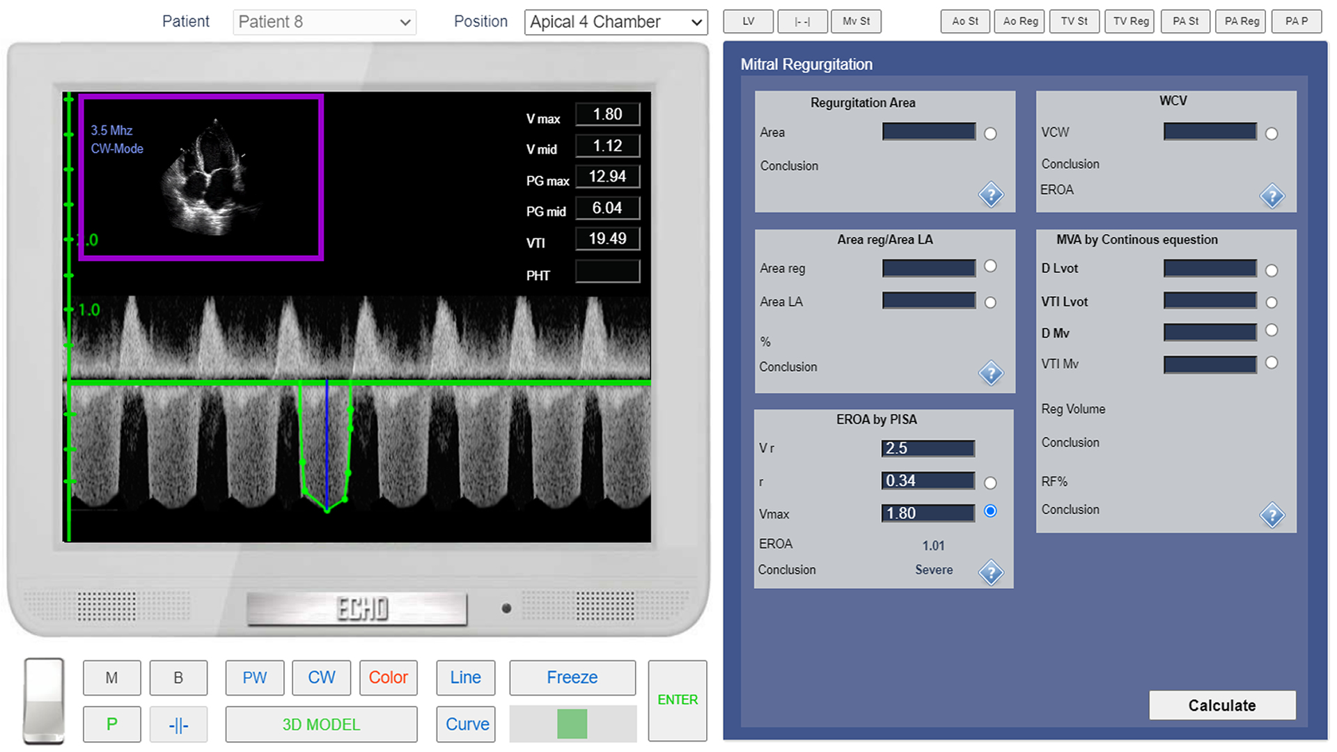
Apical 4 Chamber view, CW Doppler. Mitral Regurgitation Vmax (maximum velocity) Assessment.