Assessment degree of aortic Regurgitation by Regurgitation volume and fraction (SV method)
Echocardiography Simulator
What about a little bit of theory before beginning the simulation? >>>
- From the List <<Patients>> Choose Patient 4 (User can Choose other patients with aortic regurgitation as well).
- From the List <<Positions>> Choose Parasternal Long (Left Parasternal View, Long axis of LV). or find the same position with 3D Transducer.
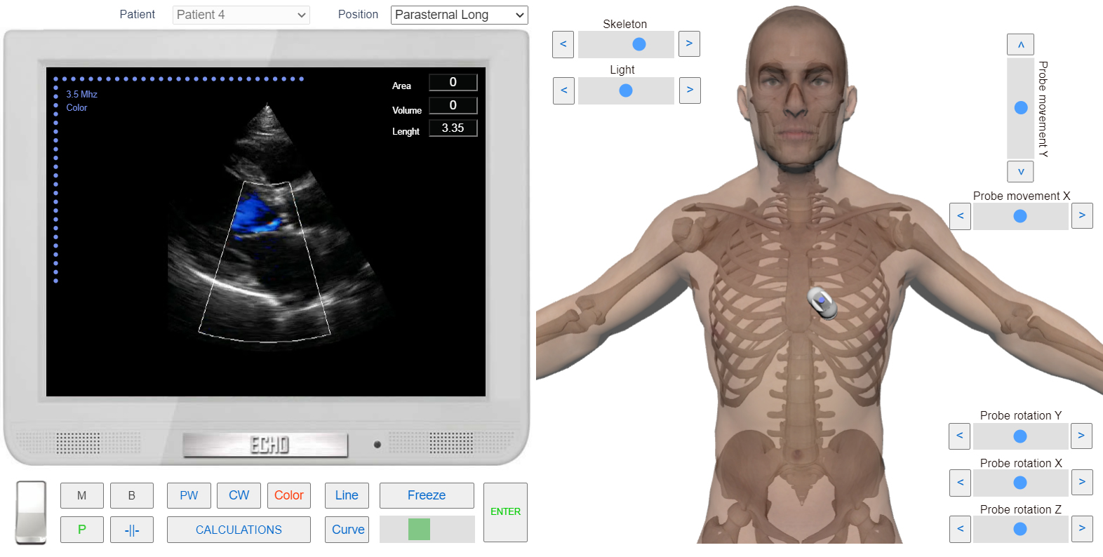
Left Parasternal View, the Long axis of LV. Color Doppler. Aortic regurgitation.
- Click the button <<Freeze>> to freeze image.
- Click Button <<Calculations>> Click Tab <<Ao Reg>> Click Radio Button <<D Lvot>>
- Click the button <<Line>> Measure linear size of Left Ventricular Outflow Tract (LVOT). See Linear measurements.
- Click the button <<Enter>>
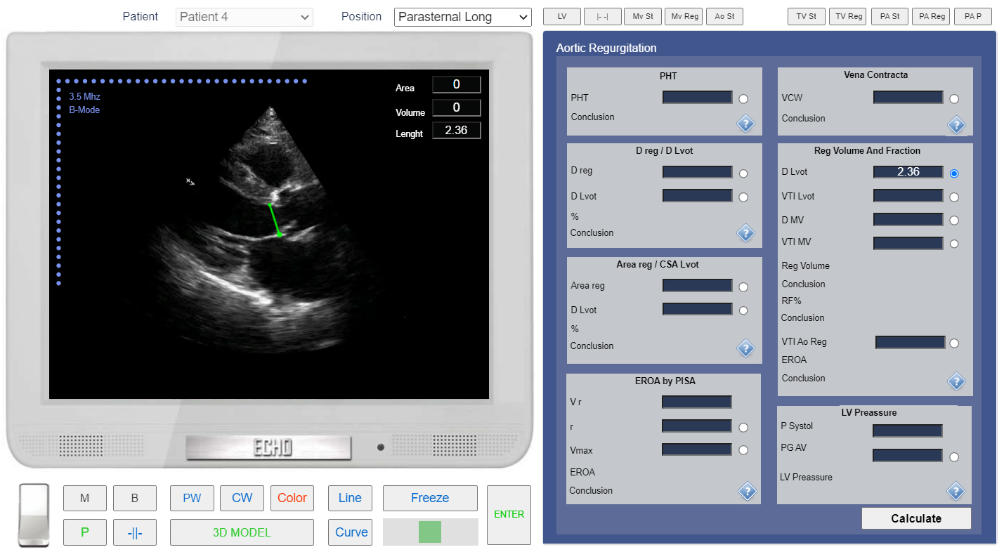
Left Parasternal View. The long axis of LV. Measurement of the Left Ventricular Outflow Tract (LVOT) diameter.
- From the List <<Positions>> Choose Apical 4 Chamber or find the same position with 3D Transducer.
- Click the button <<Freeze>> to freeze image.
- in the Tab <<Ao Reg>> Click Radio Button <<D mv>>
- Click the button <<Line>> Measure Mitral Anullus Diameter. See Linear measurements.
- Click the button <<Enter>>
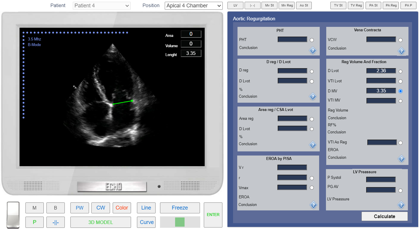
Apical 4 chamber view. Measurement of the Mitral Annulus Diameter.
- In the Tab <<Ao Reg>> Click Radio Button <<VTI mv>>
- Click the button <<PW>> to start Pulse Wave doppler examination. Place Control Volume Beetwen Mitral Valve Leaflets and Click.
- Click the button <<Curve>> place points around spectrogram of transmitral diastolic flow. See Spectral Doppler Measurements.
- Click the button <<Enter>>
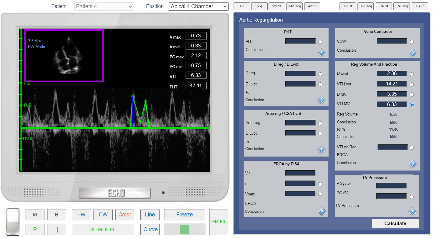
Apical 4 chamber view. PW Doppler. VTI mv measurement.
- From the List <<Positions>> Choose Apical 5 Chamber or find the same position with 3D Transducer.
- In the Tab <<Ao Reg>> Click Radio Button <<VTI Lvot >>
- Click the button <<CW>> to start Continous Wave doppler examination. Place Control Volume in LVOT and Click.
- Click the button <<Curve>> place points around spectrogram of LVOT Systolic Flow. See Spectral Doppler Measurements.
- Click the button <<Enter>>
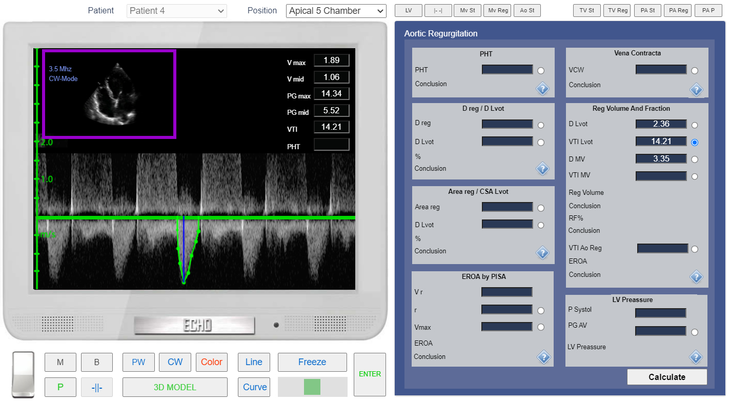
Apical 5 chamber view. CW Doppler. VTI LVOT measurement.
Let's Calculate EROA (Effective Regurgitant Orifice Area).
- In the Tab <<Ao Reg>> Click Radio Button <<VTI Ao Reg >>
- Click the button <<Curve>> place points around spectrogram of Ao regurgitation. See Spectral Doppler Measurements.
- Click the button <<Enter>>
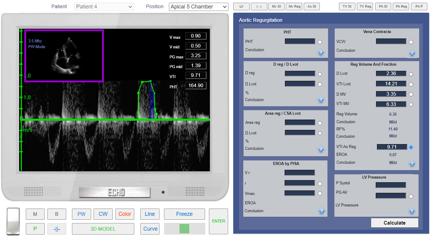
Apical 5 chamber view. CW doppler. Measurement of the VTI of Ao regurgitation.