degree of aortic Regurgitation by Regurgitation volume and fraction
Echocardiography Textbook
The volume of Aortic regurgitation is the difference between the flow in the outflow tract of the left ventricle and the transmitral flow:
![]()
RV - regurgitation volume. SVmv - transmitral stroke Volume. SVLVOT - LVOT Stroke Volume.
To calculate the transmitral Stroke Volume (SV), it is necessary to measure the D-diameter of the mitral annulus and VTI of the transmitral flow.
![]()
D - diameter of the mitral annulus. VTIMV - VTI of the transmitral flow.
To calculate the stroke volume through the outflow tract of the left ventricle, it is necessary to measure the D-diameter of the LVOT and the VTI of the flow in the outflow tract of the left ventricle (LVOT).
![]()
D - diameter of the LVOT. VTILVOT - VTI of the LVOT flow.
- LVOT diameter measured at the annulus in systole and pulsed Doppler from apical views at same site.
- Mitral annulus measured at middiastole; pulsed Doppler at the annulus level in diastole.
- Total LV SV can also be measured by the difference between LV end-diastolic volume and endsystolic volume.
- LV volumes are best measured by 3D. Contrast may be needed to better trace endocardial borders. If 3D not feasible, use 2D method of disks.
Assessment of Aortic regurgitation by the regurgitation volume:
- Mild < 30
- Mild-Moderate 30 - 44
- Moderate-severe 45 - 59
- Severe > 60
Regurgitation fraction (RF%) is determined by the following formula:
![]()
Assessment of Aortic regurgitation by the regurgitation fraction (RF%):
- Mild < 30
- Mild-Moderate 30 - 39
- Moderate-severe 40 - 49
- Severe > 50
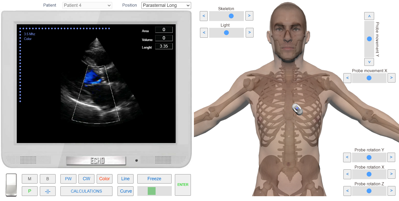
Left Parasternal View, the Long axis of LV. Color Doppler. Aortic regurgitation. Simulation By Echocardiography Online Simulator MyEchocardiography.com
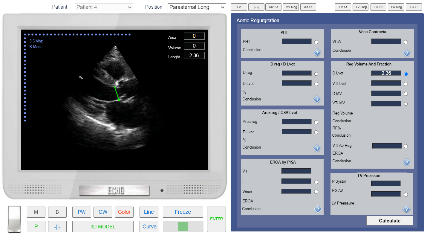
Left Parasternal View. The long axis of LV. Measurement of the Left Ventricular Outflow Tract (LVOT) diameter. Simulation By Echocardiography Online Simulator MyEchocardiography.com
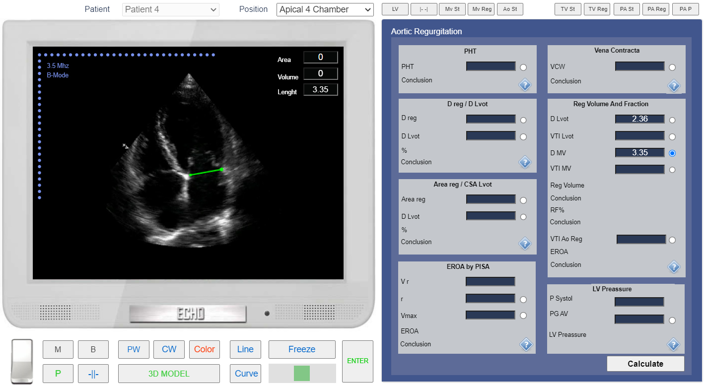
Apical 4 chamber view. Measurement of the Mitral Annulus Diameter. Simulation By Echocardiography Online Simulator MyEchocardiography.com
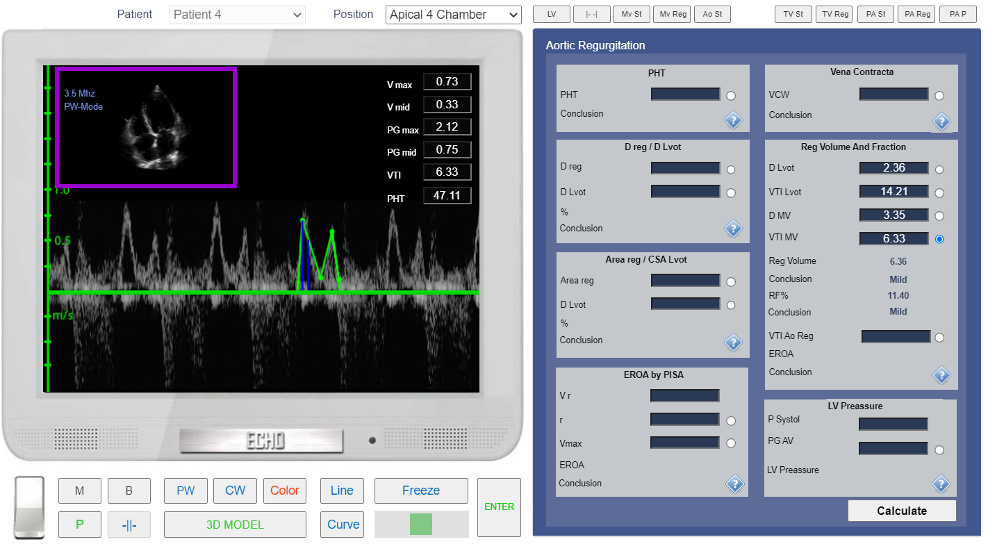
Apical 4 chamber view. PW Doppler. VTI mv measurement. Simulation By Echocardiography Online Simulator MyEchocardiography.com
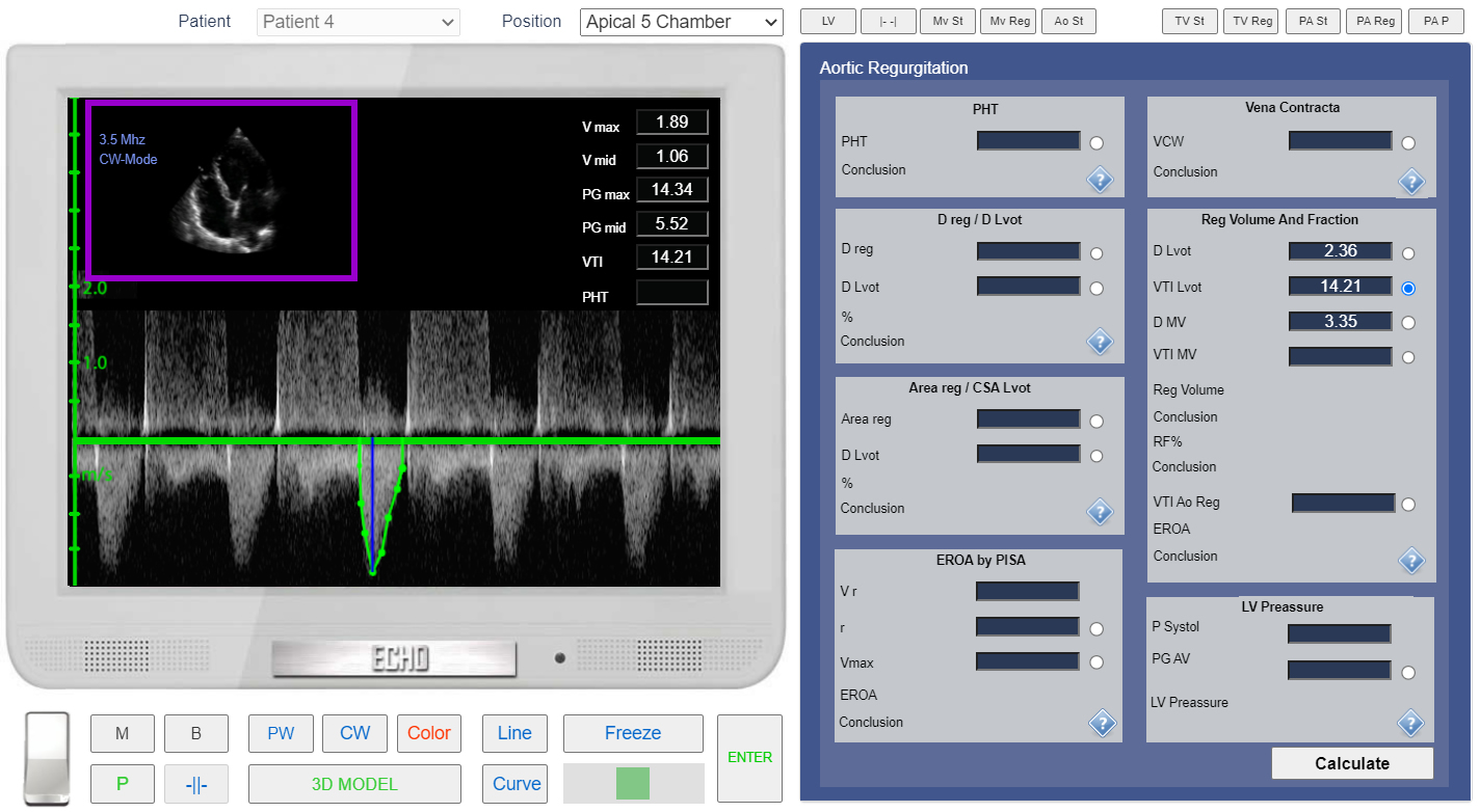
Apical 5 chamber view. CW Doppler. VTI LVOT measurement. Simulation By Echocardiography Online Simulator MyEchocardiography.com
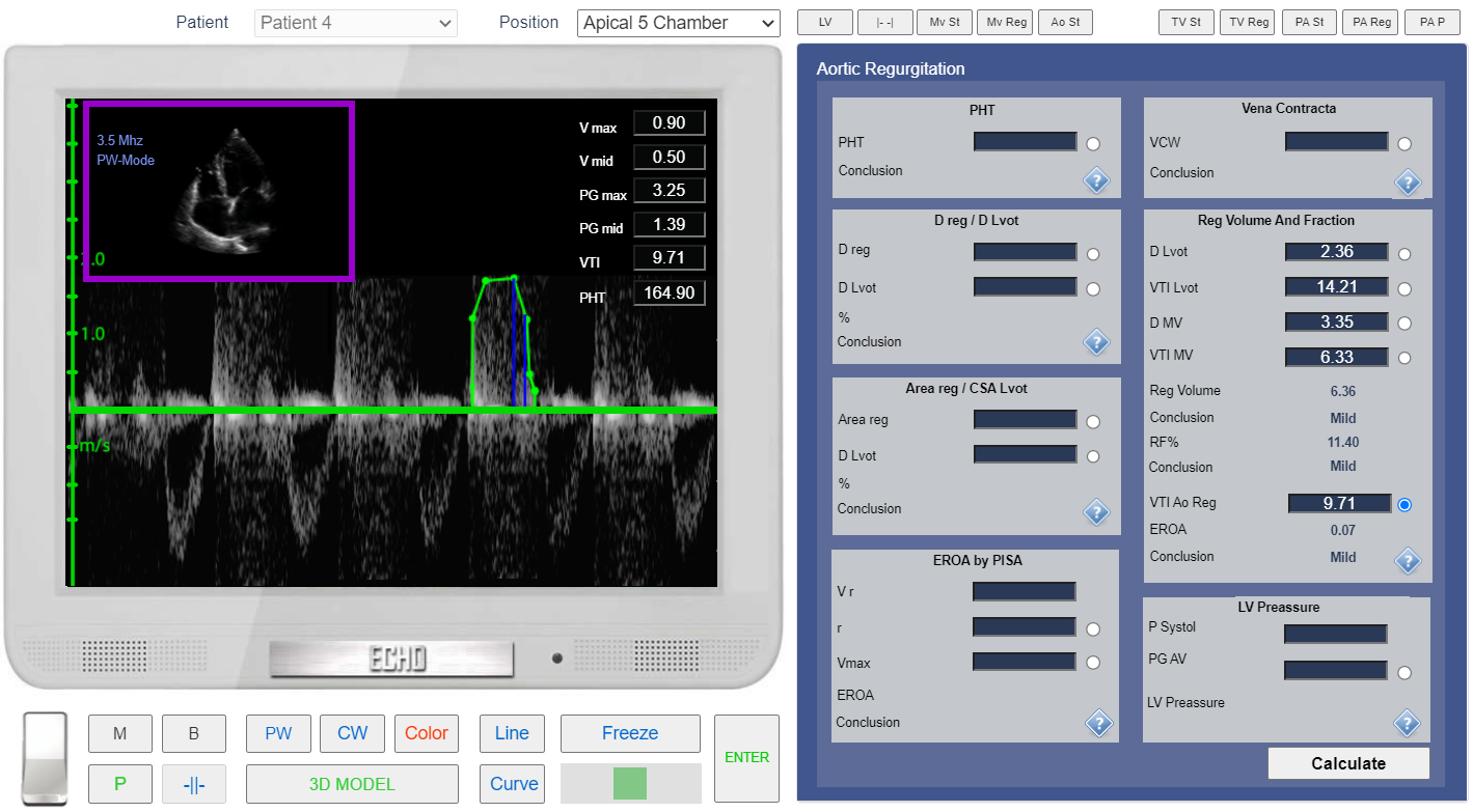
Apical 5 chamber view. CW doppler. Measurement of the VTI of Ao regurgitation. Simulation By Echocardiography Online Simulator MyEchocardiography.com
ASE Guideline - Recommendations for Noninvasive Evaluation of Native Valvular Regurgitation