Echocardiographic evidence of aortic regurgitation
Echocardiography Textbook
One and two dimensional Echocardiography
The only relatively reliable specific finding can be Diastolic vibration of the anterior leaflet of the mitral valve is caused by a reversed turbulent outflow of blood from the aorta into the left ventricle (one-dimensional echocardiography).
Non-specific symptom: LV Myocardial hypertrophy and dilatation.
Color Doppler
The most informative method is color doppler. The best positions for evaluation: Apical 5 Chamber view and the Left Parasternal View, the Long axis of the heart. Regurgitation flow is observed in the diastolic phase, and returns to the left ventricle. If examining from Apical 5 Chamber View, The flow is directed towards the transducer accordingly encoded in red color.
When examining from a parasternal approach, the long axis of the heart, the regurgitation flow can be directed towards or away from the transducer. This depends on the orientation of the outflow tract to the ultrasound beam. If the LV outflow tract is in front of the transducer, then the regurgitation flow is directed away the transducer and is accordingly coded in blue color. When positioned at the back, the flow is directed towards the transducer and is coded in red color.
Regurgitation size and volume depend on the severity of regurgitation.
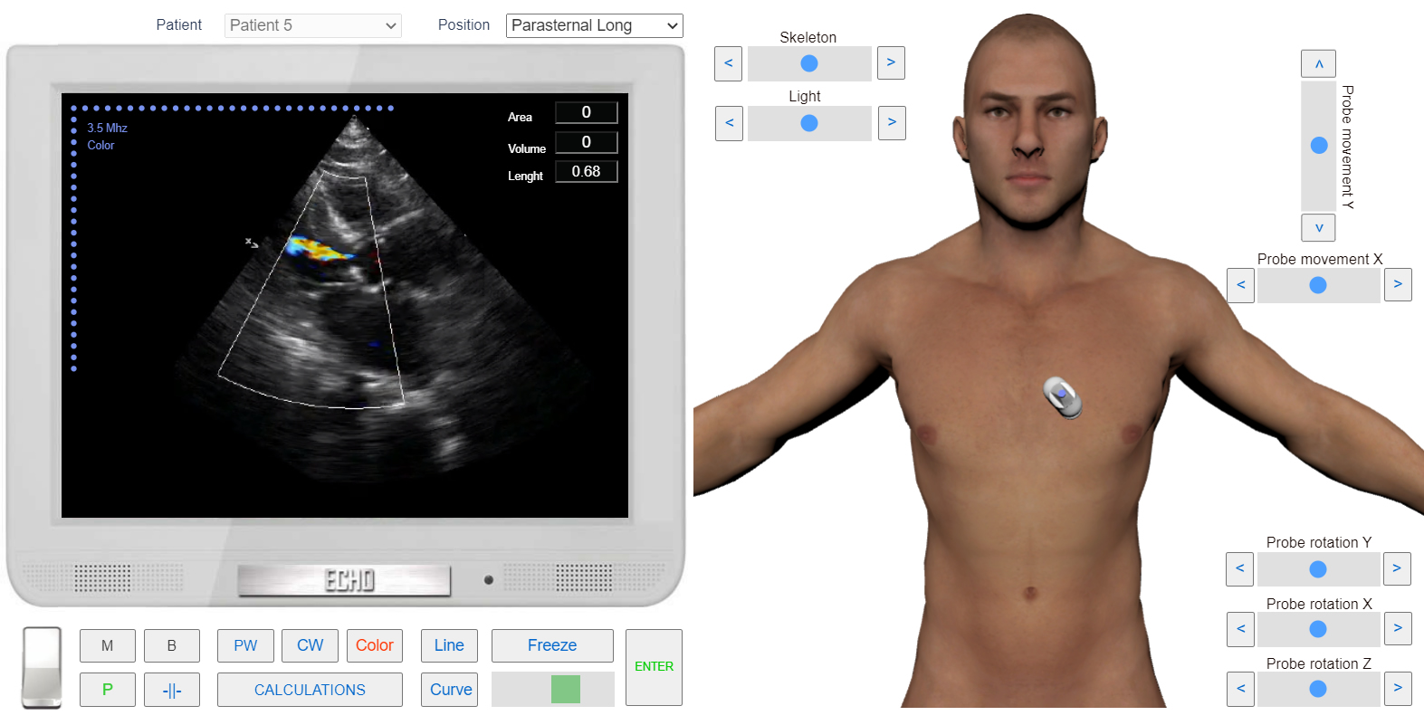
Left Parasternal View, the long axis. Color Doppler. Aortic regurgitation. Simulation by Echocardiography Online Simulator MyEchocardiography.com
Spectral Doppler (PW, CW)
The best positions for evaluation: Apical 5 Chamber View.
To find the regurgitation and determine the depth of its penetration the sample volume is placed in the outflow tract of the left ventricle, at the level of the junction of the aortic valve leaflets. After it has to be moved at different distances from the Aortic valve (in LV).
The flow of regurgitation is clearly visible on the spectrogram (both in PW and CW mode) as a characteristic spectrum that is abowe the baseline. The flow captures the entire diastole (from the closing of the aortic valve to the opening of the mitral valve).
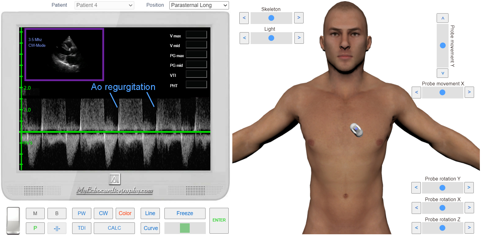
CW Doppler. Aortic regurgitation. Simulation by Echocardiography Online Simulator MyEchocardiography.com
Methods of assessment severity of the Aortic Regurgitation:
- PHT
- Regurgitation jet width / LVOT diameter
- Regurgitation jet area / CSA LVOT
- Vena Contracta
- Regurgitation volume and fraction (SV method)