Echocardiographic evidence of mitral stenosis
Echocardiography Textbook
One Dimensional Echocardiography (M-Mode)
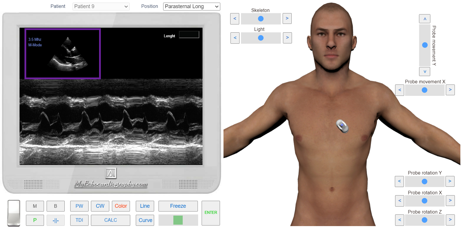
- Decrease in the rate of diastolic closure of the anterior leaflet of the mitral valve.
- Unilateral movement of the mitral valve anterior and posterior leaflets.
Due to the high pressure in the left atrium during diastole, the valve leaflets remain open and, unlike the norm, do not close after the left ventricle early rapid filling.
The curve of the mitral valve movement has a п-shaped form instead of an M-shaped.
The unilateral movement of the anterior and posterior leaflets of the mitral valve can be seen, which is due to the fusion of the leaflets. (Normally, the leaflets move in different directions).
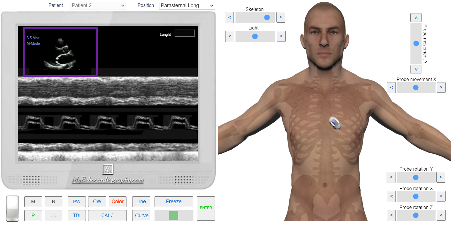
Left parasternal view, the lond axis of the heart. One dimensional Echocardiography (m-mode). Normal Mitral Valve. Simulation by Echocardiography Online Simulator MyEchocardiography.com
Left parasternal view, the lond axis of the heart. One dimensional Echocardiography (m-mode). Mitral Stenosis. Decrease in the rate of diastolic closure of the anterior leaflet of the mitral valve and Unilateral movement of the mitral valve anterior and posterior leaflets. Simulation by Echocardiography Online Simulator MyEchocardiography.com
Two Dimensional Echocardiography (B-Mode)
From the early stages of mitral stenosis, in the diastole phase, there is a dome-shaped bulging of the anterior leaflet of the mitral valve (towards the septum) which is caused by an increase in pressure on its unfixed part (Left parasternal view, the lond axis of the heart).
In the later stages of mitral stenosis, the valve leaflets thicken and become rigid. Fusion of the leaflets and restriction of movement are often observed. From the left parasternal position, the short axis of the heart, there is a decrease in the area of the mitral orifice, which takes the form of an ellipsoid or gap. There is an increase in the size of the left atrium.
An increase in pressure in the pulmonary circulation causes hypertrophy and dilatation of the right heart. The hepatic veins and inferior vena cava can be dilated.
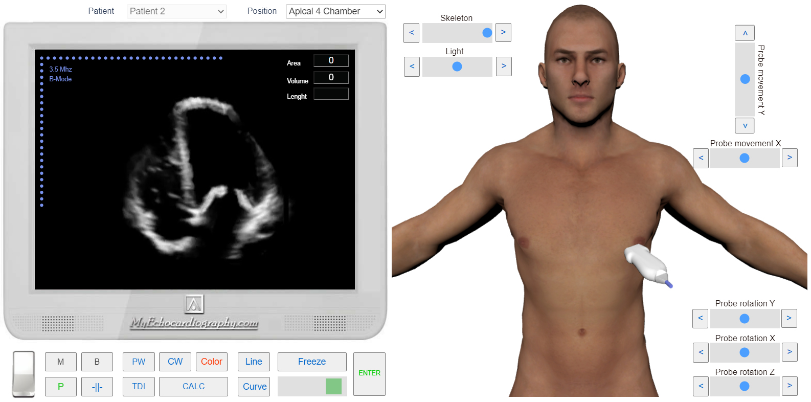
Apical 4 chamber view. 2D Echocardiography. Mitral Stenosis. Thick and rigid mitral valve leaflets. Simulation by Echocardiography Online Simulator MyEchocardiography.com
Spectral Doppler (PW, CW)
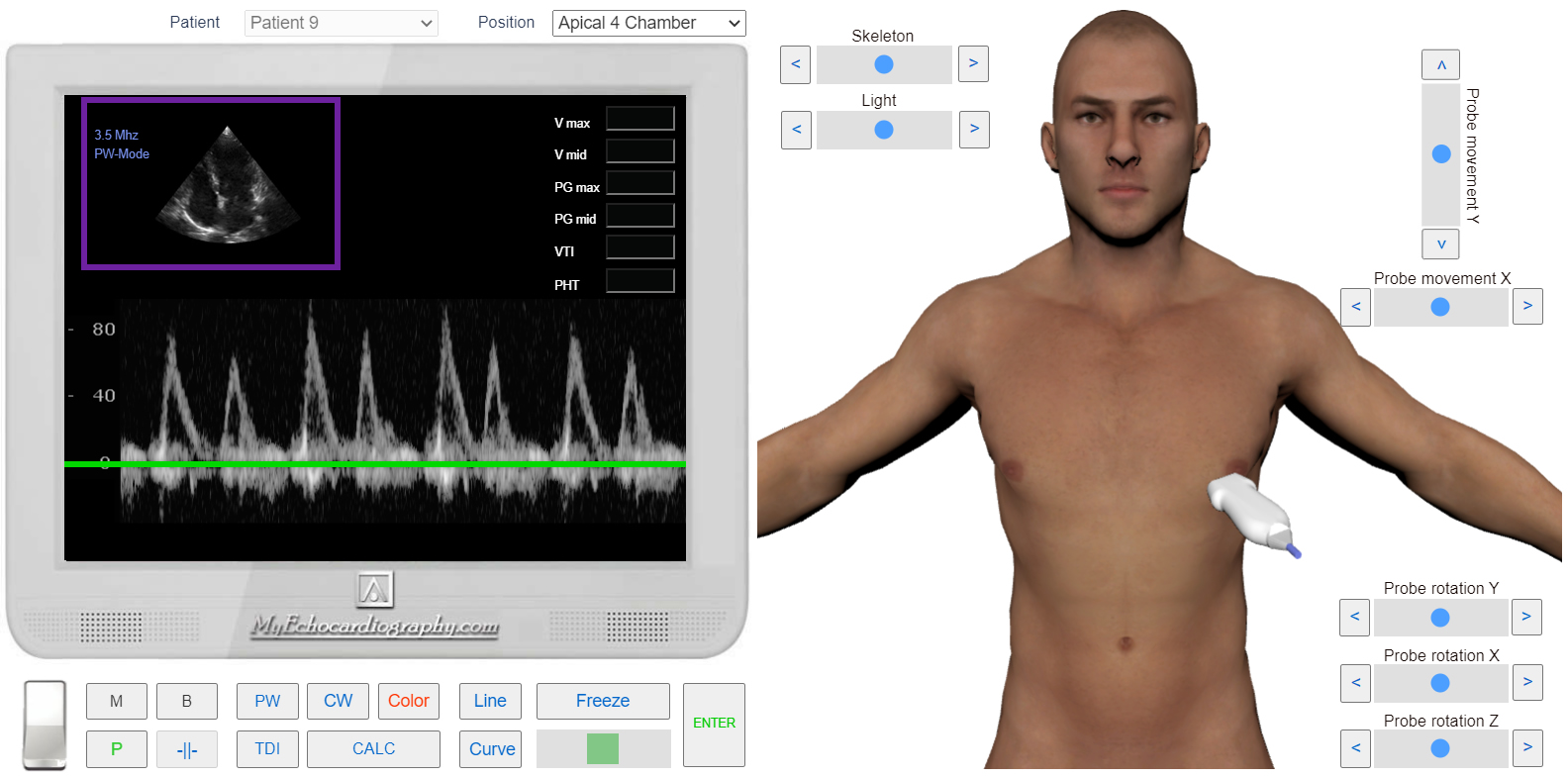
- Increase in velocity of early transmitral diastolic flow - that caused by the increase of pressure gradient between LA and LV.
- Spectrogram extension - that caused by a reduction of the fall of the diastolic filling rate.
Apical 4 Chamber View. PW Doppler. Normal transmitral Diastolic Flow. Simulation by Echocardiography Online Simulator MyEchocardiography.com
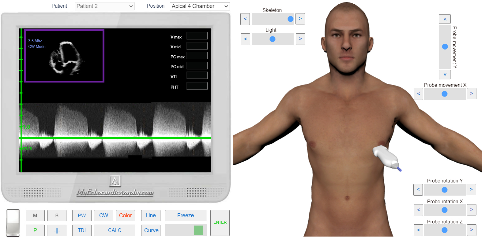
Apical 4 Chamber View. CW Doppler. Mitral stenosis. Increase in velocity of early transmitral diastolic flow and spectrogram extension. Simulation by Echocardiography Online Simulator MyEchocardiography.com
Methods of measurement of the Mitral Valve Area (MVA)
- MVA Tracing
- PISA
- PHT
- Mitral Valve Area (MVA) by Continuity Equation
- Mean Pressure Gradient (PG mean)