Dilated cardiomyopathy
Echocardiography Textbook
Dilated cardiomyopathy is characterized by significant dilatation of heart chambers (mainly left ventricle) and systolic dysfunction. Despite the different etiologies, the 2D echocardiographic picture is the same in all cases. The left ventricle is spherical in shape (linear dimensions on its short and long axis are approximately the same. The ratio is close to 1).
Quantitative values of all systolic heart function characteristics are reduced (ejection fraction <30%, stroke volume, cardiac output, etc. are reduced). There are rare cases when the dilatation is minimal and the left ventricular dysfunction is significant.
Tthickness of the wall of the left ventricle is mostly within the norm, in some cases its thinning or thickening may be detected. Myocardium mass is increased in all cases. Dilatation of the chambers and fibrous rings leads to the development of a relative deficiency of the valves (mainly mitral regurgitation. tricuspid regurgitation is quite common). Regurgitation can be revealed by color and spectral doppler.
Signs of left ventricular diastolic dysfunction are also not uncommon. The presence of diastolic dysfunction is a worse prognostic sign.
In the dilated akinetic-hypokinetic left ventricular cavity, are conditions for thrombus formation.
On the one-dimensional echocardiogram of the mitral valve, we can see increased E-point septal separation (increases the distance from the E-point to the septum. Normally it is up to 5 mm).
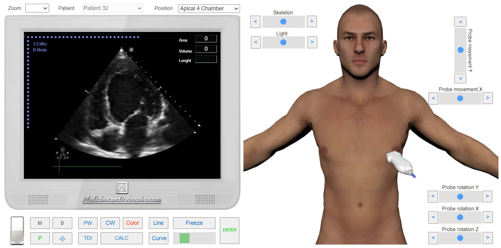
Apical 4 Chamber View. Dilated Cardyomiopathy. Simulation By Echocardiography Online Simulator MyEchocardiography.com
Simulate Dilated Cardiomyopathy >>>