pericardial effusion
Echocardiography Textbook
In the case of Pericardial Effusion the volume of the liquid is greater than its physiological value (Trivial 50-80 ml). Echocardiography is the initial procedure of choice to detect the presence of a Pericardial Effusion because it can be performed with minimal delay and has an accuracy of nearly 100%.
One Dimensional Echocardiography (M-mode) shows the persistence of an echo-free space between the epicardium and parietal pericardium throughout the cardiac cycle. separation of the two layers that is seen only in systole represents a normal or clinically insignificant amount of pericardial fluid (trivial PE), whereas a separation that is present in both systole and diastole is associated with effusions of >50 mL (small PE).
M-modal examination is best done from a parasternal approach.
Normally, the parietal layer of the pericardium is one of the brightest structures of the heart. If the signal gain is maximally reduced only the parietal pericardium will remain on the screen because it reflects the ultrasonic beam more than other structures of the heart.
Normally, the layers of the pericardium move in parallel. With exudative pericarditis, the parallel movement disappears, we see the separation of the layers and the formation of an echo-negative space between them. Diagnostic value has diastolic separation of the layers.
Movement of the parietal pericardium is reduced or completely disappears.
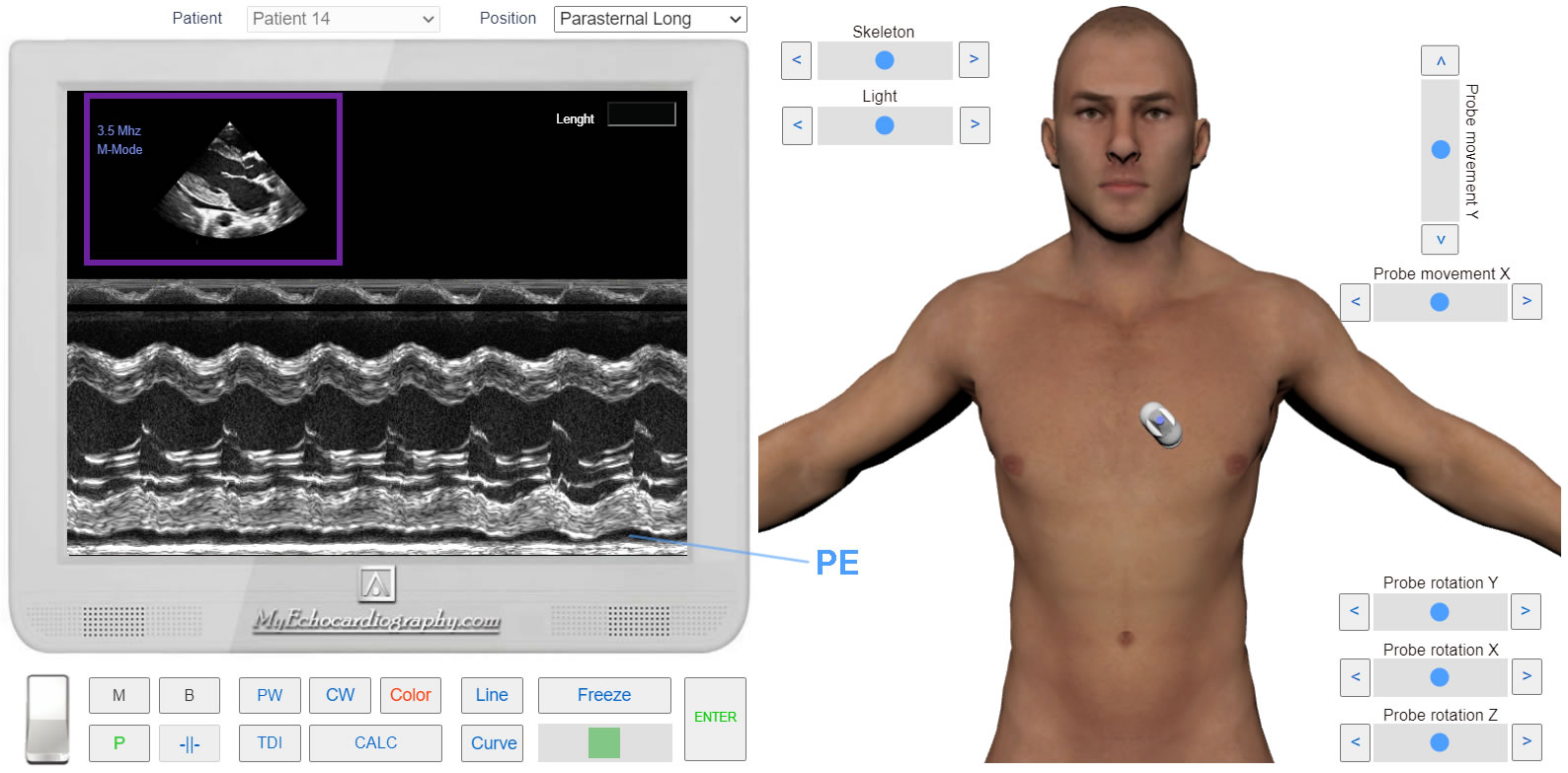
M-modal examination. Left Parasternal View, Long axis of the Heart. Pericardial effusion. Simulation By Echocardiography Online Simulator MyEchocardiography.com
The main diagnostic method - is Two-Dimensional Echocardiography (B-mode).
It helps to identify pathological fluid, determine the volume and its effect on hemodynamics.
Studies are mainly carried out from the left Parasternal approach (long and short axis of the heart) from the Apical four-chamber position and the Subcostal approach.
When examined from the Left Parasternal approach, long axis, fluid is most often located behind the posterior wall of the left ventricle.
Four pulmonary veins are attached to the left atrium, the layers of the pericardium of this area are fused and do not separate even with the accumulation of a large volume of fluid. Therefore, in this area, the accumulation of fluid is very rare, but sometimes it is observed.
When examined from the Apical Four-chamber position, the fluid is mostly behind the wall of the right atrium and right ventricle.
2D echocardiography shows the persistence of echo-free space between the layers of pericardium. If the age of accumulation of fluid is 1 month, then it is transparent, if more than 1 month, fibrin strands are formed. The more pronounced fibrin, the long process we are dealing with.
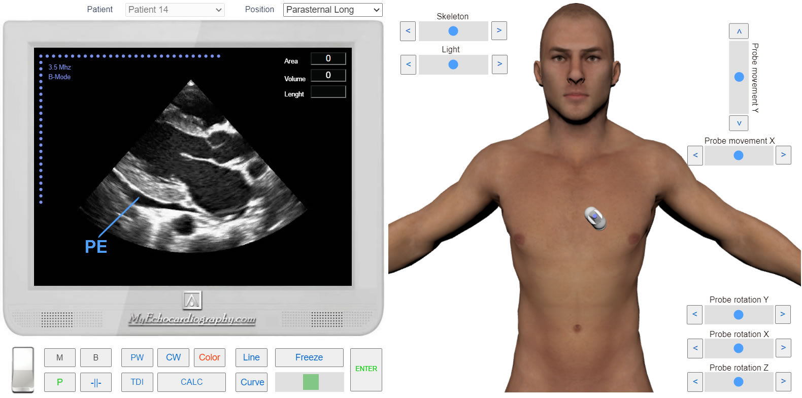
Left Parasternal View, Long axis of the Heart. Pericardial effusion. Simulation By Echocardiography Online Simulator MyEchocardiography.com
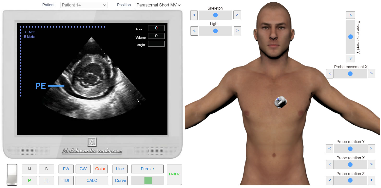
Left Parasternal View, Short axis at the level of the mitral valve. Pericardial effusion. Simulation By Echocardiography Online Simulator MyEchocardiography.com
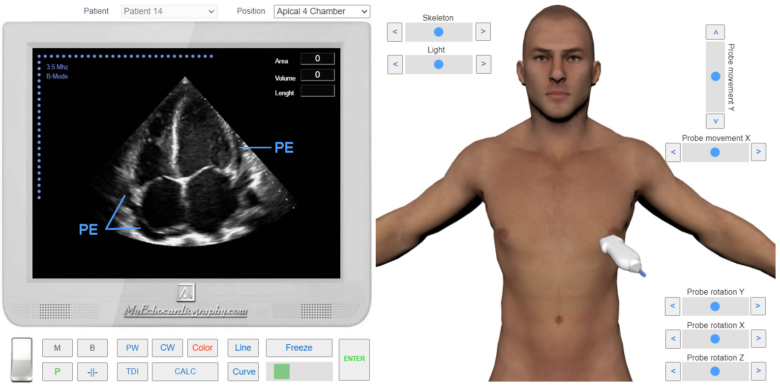
Apical 4 Chamber View. Pericardial effusion. Simulation By Echocardiography Online Simulator MyEchocardiography.com
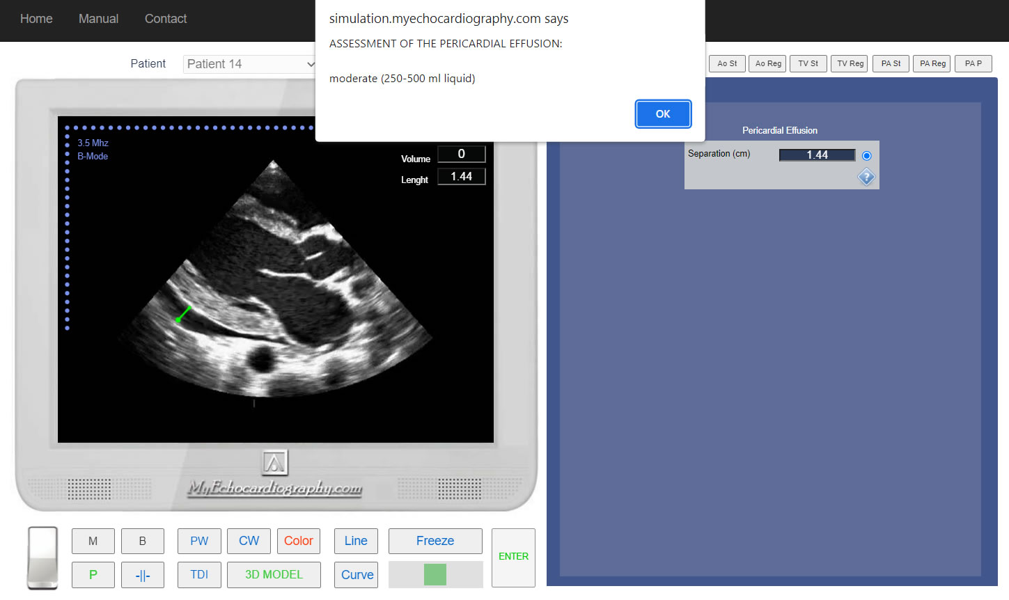
Determining the volume of Pericardial Effusion
- Trivial (seen only in systole).
- 5-10 mm separation - small (100-250 ml liquid).
- 10-20 mm separation - moderate (250-500 ml liquid).
- >20 mm separation - large (more than 500 ml of liquid).
- >25 mm - very large
Left Parasternal View, Long axis of the Heart. Assessment of the pericardial effusion. Simulation By Echocardiography Online Simulator MyEchocardiography.com
ASSESSMENT OF THE PERICARDIAL EFFUSION. Simulation By Echocardiography Online Simulator MyEchocardiography.com
ASE Guideline for Imaging of Patients with Pericardial Disease
How to Simulate pericardial effusion and its assessment?