Right ventricular outflow tract (RVOT) flow spectral doppler spectral (PW, CW) doppler
Echocardiography textbook
The study is better to perform from a Parasternal Approach, short axis of the heart, at the level of the Aortic Valve or Pulmonary Artery. The Sample Volume is placed under the cusps of the pulmonary valve, towards the right ventricle (1 cm proximally).
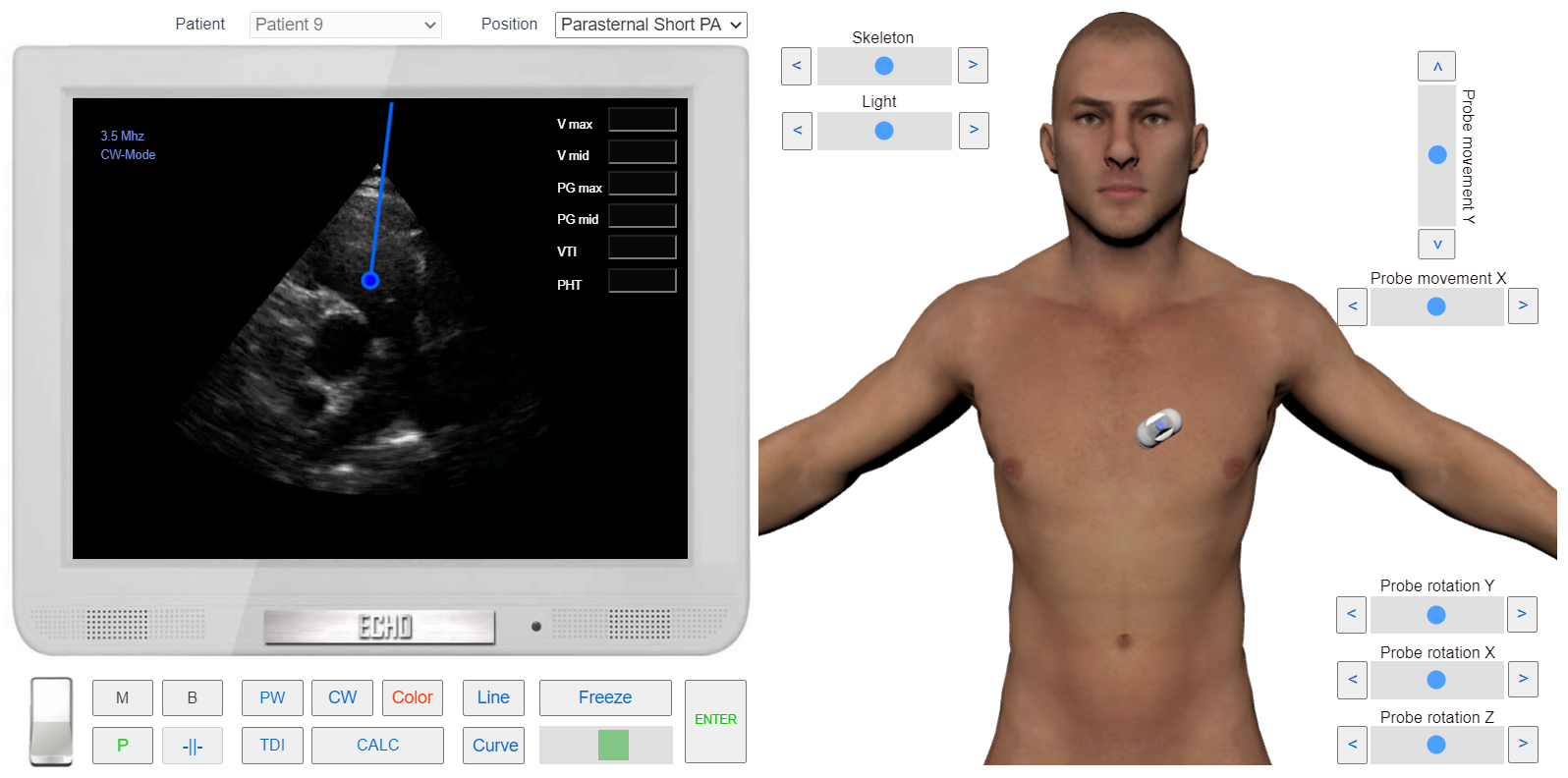
RVOT Flow examination. Sample Volume position. Parasternal view, Short axis of the heart at the level of Pulmonary Artery
Flow occurs in the diastole phase and is directed from the right ventricle to the pulmonary artery, away from the transducer, and located below the Baseline. The flow, in contrast to the aortic, has a small amplitude. In 50-65% of cases, physiological regurgitation is observed on the pulmonary valve.
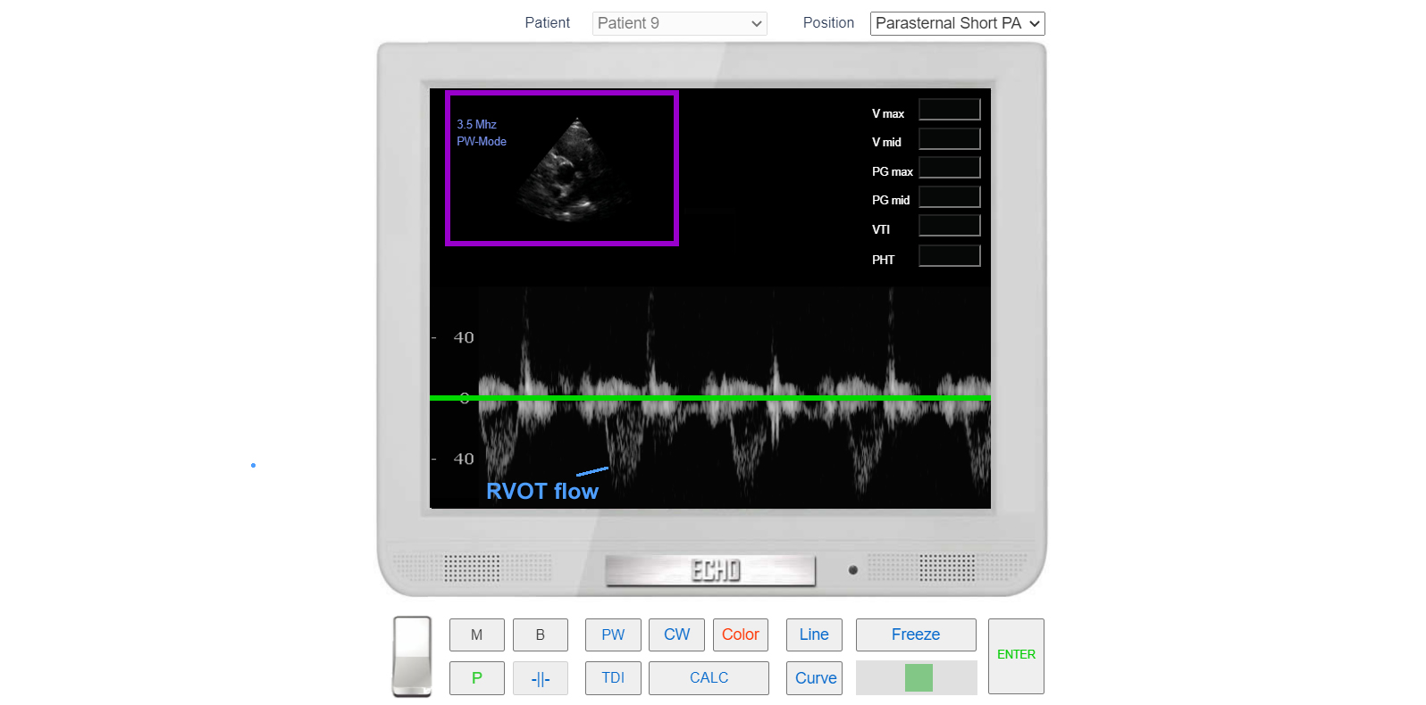
RVOT flow. PW Doppler