IVC and hepatic veins flow spectral (PW, CW) doppler
Echocardiography textbook
The study is carried out from a Subcostal approach, the long axis of the Inferior Vena Cava. The control volume is placed in IVC or in the hepatic vein 1-2 cm proximally from its junction with the inferior vena cava.
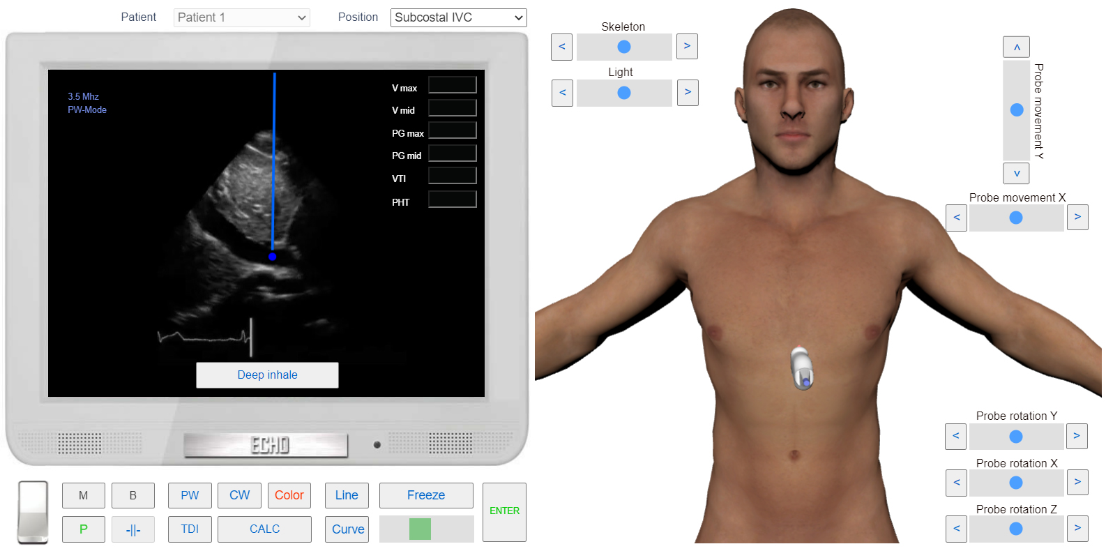
IVC flow examination. Sample Volume position. Subcostal IVC View.
Normally, the flow is observed both in the systole and diastole phases. Consists of a Systolic, Diastolic, and Atrial Component.
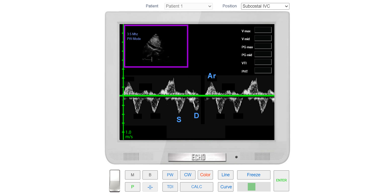
IVC flow. PW Doppler