one-dimensional echocardiography
Echocardiography Textbook
The one-dimensional examination is mainly carried out from the parasternal position, along the long axis of the heart. The angle of inclination of the m-modal cursor is chosen so that the ultrasound beam crosses the mitral valve, aortic valve, and left ventricle.
Nowadays, one-dimensional echocardiography is mainly used as an additional method for taking measurements. The cursor should be positioned strictly perpendicular to the structures of the heart, in this case, measurements can be made with great accuracy.
The typical advantage of M-modal echocardiography is its high resolution. Therefore, a one-dimensional echocardiogram provides significant information from rapidly moving structures. For example vibration of the anterior mitral leaflet cannot be detected on a 2D echocardiogram.
The advantage of two-dimensional echocardiography is its two-dimensional nature. One-dimensional echocardiography does not provide spatial information.
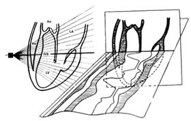
The relationship between one-dimensional and two-dimensional echocardiography.
On the M-modal graph, distances are located vertically, time is horizontal. The graph gives the following information:
- Time - on the horizontal axis.
- Distance - on the vertical axis.
- Saturation (density) of the ultrasound signal - on the display the structure with different brightness (echogenicity).
One-dimensional (M-Mode) Examinations
- M-modal examination of the mitral valve.
- M-modal examination of the aorta and aortic valve.
- M-modal examination of the left ventricle.
How to Simulate one-dimensional echocardiography (M-mode)?