Tissue Doppler
Echocardiography Textbook
The myocardium has subendocardial and epicardial layers, with the former having longitudinally arranged myofibres. During ventricular contraction, various layers exert varying tension with the endocardium moving greater distances. Tissue Doppler imaging examines the longitudinal component of myocardial contraction throughout the cardiac cycle.
Tissue Doppler imaging is obtained using pulsed wave tissue Doppler or colour tissue Doppler imaging (CTDI).
Pulsed wave TDI measures peak longitudinal myocardial velocity from a single segment, but has to be performed ‘on line’.
Colour tissue Doppler imaging is performed ‘off line’, and can interrogate velocities from multiple sites simultaneously.
The major disadvantage of TDI is its angle dependence i.e. if the angle of incidence exceeds 15 degrees, there is 4% underestimation of velocity. Accurate TDI imaging additionally requires high frame rates (>100fps) for image acquisition with excellent temporal resolution.
The TDI signal over a cardiac cycle has three peaks, a positive systolic peak and two negative diastolic peaks. The positive systolic wave (s’ velocity, Sa or Sm) represents myocardial contraction.
The negative waves represent the early diastolic myocardial relaxation (e’ velocity, Ea or Em) and active atrial contraction in late diastole (a’ velocity, Aa or Am)
Pulsed wave TDI velocity measurements are obtained by placing the sample volume at the mitral annular level (denoted Sa/s’ or Ea/e’ or Aa/a’) or within the basal LV myocardial segment (denoted Sm or Em or Am).
Tissue Doppler imaging velocities can be measured either from the septal or lateral annulus, but the current recommendation is that e’ velocity is expressed as the average of septal and lateral measurements.
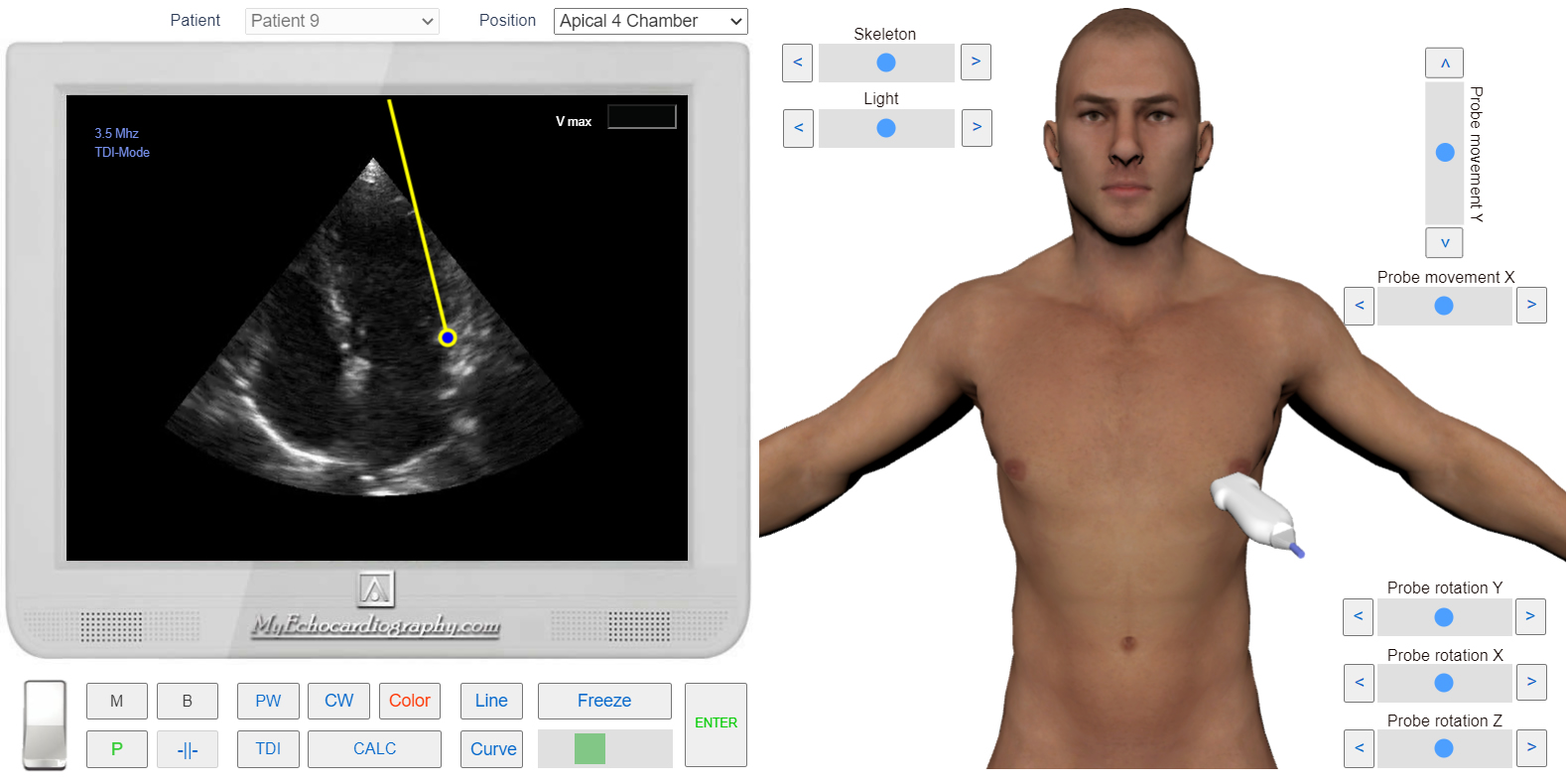
Echocardiography Online Simulator MyEchocardiography.com allows simulation of Pulsed wave TDI in Apical 4 Chamber View at the mitral annular level (Septal and Lateral).

Apical 4 chamber view. Sample Volume position for the Septal TDI. Simulation by Echocardiography Online Simulator MyEchocardiography.com
Apical 4 chamber view. Sample Volume position for the Lateral TDI. Simulation by Echocardiography Online Simulator MyEchocardiography.com

Tissue Doppler (TDI). S' - The positive systolic wave represents myocardial contraction. e' - early diastolic myocardial relaxation. a' - active atrial contraction in late diastole. Simulation by Echocardiography Online Simulator MyEchocardiography.com
How to Simulate Tissue Doppler examination? >>>