LOCAL SYSTOLIC function
Echocardiography Textbook
The American Association of Echocardiography (ASE) recommends using a 16-segment model to assess regional myocardial contractility based on the following:
- Anatomical logic.
- Easy identification of each segment with anatomical boundaries.
- The connection of each segment to the blood supply from a particular coronary artery.
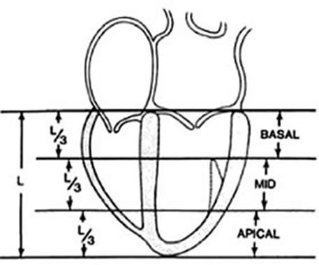
This model divides the myocardium into three levels and 16 segments. When dividing into levels, the papillary muscles are considered the anatomical border.
- Basal level (Basal) - from the mitral valve's fibrous ring to the papillary muscles' end.
- Middle level (Mid) - from the end of the papillary muscles to its beginning.
- Apical level - from the beginning of the papillary muscles to the top of the heart.

Division of the left ventricle into basal, middle, and apical levels. Left parasternal position, long axis.
Division of the left ventricle into basal, middle, and apical levels. Apical 4 chamber view.
The basal and middle levels are divided into six equal segments, and the apical level is divided into four segments.
It is possible to classify each segment according to the blood supply of 3 coronary arteries (We should consider differences between the individuals in the coronary blood supply).

Division of myocardium into 16 segments for two-dimensional echocardiography. 1. Left parasternal view, long axis. 2. Apical four-chamber view. 3. Apical two-chamber view. 4. Left parasternal view, short axis, at the level of the mitral valve. 5. Left parasternal view, short axis, at the level of the papillary muscles. 6. Apex.
The regional contractility assessment system is based on the assessment of each segment.
Recommended quantitative criteria for the assessment of regional contractility:
- Normal contractility - systolic increase in ventricular wall thickness by more than 50% (1 point).
- Hypokinesia - systolic increase in ventricular wall thickness less than 40% (2 points).
- Akinesia - systolic increase in ventricular wall thickness of less than 10% (3 points).
- Dyskinesia - depression on the opposite side of the wall during systole and its thinning (4 points).
- Aneurysm - concavation on the opposite side of the wall during systole, dilatation, systolic thinning, and diastolic deformation remains during the diastole phase (5 points).
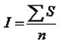
The following formula calculates the regional contractility index:
How to Simulate Assessment of the Local Systolic Function? >>>